Is Kaempferol a potential height increase supplement?

Kaempferol levels were elevated in one study after administration of fructus sophorae extract.  Kaempferol is found in foods like strawberries, grapes, apples, etc.

It is important to note that in the following study ATDC5 cells were involved.  They are not like mesenchymal stem cells in that they are already chondrocyte progenitor cells and are primed for chondrogenesis.

Kaempferol Induces Chondrogenesis in ATDC5 Cells through Activation of ERK/BMP-2 Signaling Pathway.

“Endochondral bone formation occurs when mesenchymal cells condense to differentiate into chondrocytes, the primary cell types of cartilage. We investigated whether kaempferol induces chondrogenic differentiation in clonal mouse chondrogenic ATDC5 cells. Kaempferol treatment stimulated the accumulation of cartilage nodules in a dose-dependent manner. Kaempferol-treated ATDC5 cells stained more intensely with alcian blue staining than control cells, suggesting greater synthesis of matrix proteoglycans in the kaempferol-treated cells. Kaempferol induced greater activation of alkaline phosphatase activity than control cells, and it enhanced the expression of chondrogenic marker genes, such as collagen type I, collagen type X, OCN, Runx2, and Sox9{these are a lot of osteogenic genes too}. Kaempferol induced an acute activation of extracellular signal-regulated kinase (ERK) but not c-jun N-terminal kinase or p38 MAP kinase. PD98059, an inhibitor of MAPK/ERK, decreased in stained cells treated with kaempferol. Furthermore, kaempferol greatly expressed the protein and mRNA levels of BMP-2, suggesting chondrogenesis was stimulated via a BMP-2 pathway. Kaempferol has chondromodulating effects via an ERK/BMP-2 signaling pathway and could potentially be used as a therapeutic agent for bone growth disorders.

Perhaps Kaempferol could be useful for people with existing growth plates as they already have chondroprogenitor cells.

“Cells were treated with kaempferol or insulin for 21 days. ATDC5 cells treated with insulin showed chondrogenic differentiation 7 days after treatment through the condensation of stage cartilage nodules. ATDC5 cells treated with 5 μM of kaempferol showed differentiation similar to the insulin-treated cells, including the development of cartilage nodules “<-So ATDC5 cells are already primed for chondrogenesis with chondroinduction occurring merely with insulin.  Kaempferol however was more chondroinductive than mere insulin.

“phosphorylation of JNK and P38 Kinase did not occur with insulin treatmen. Treatment with 5 μM of kaempferol showed acute activation of ERK kinase after 1.5 h and the phosphorylation of P-38 Kinase was also observed from 1.5 h to 6 h. Similarly, 5 μg/ml of insulin treatment showed activation of ERK kinase and in a similar pattern to kaempferol, suggesting kaempferol has the ability to induce chondrogenic effect via the activation of ERK and P-38 MAPK. Moreover activation of ERK Kinase by Kaempferol and Insulin suggest that Kaempferol can mimic the effects of insulin with regards to activating phosphorylation of the ERK MAP kinase.”<-The phosphorylation of p38 kinase may be responsible for the enhanced chondroinductive effects and any compound that results in the phosphorylation of p38 could have similar chondrostimulatory effects.

“kaempferol affects the synthesis of matrix proteoglycans and the activity of ALP.”

“kaempferol could potentially be used to treat a variety of skeletal diseases, such as dwarfism”<-The authors have more faith in kaempferol than would be inferred from the results of the study.

Perhaps the authors are alluding to the reduction of insulin and replacement of more sources of kaempferol to grow taller during development.  More apples and less bread for taller infants.

There’s no indication that kaempferol could be chondroinductive on adult Mesenchymal Stem Cells however but it could be synergestic with other adult height increase stimulants.

Here’s a patent related to kaempferol:

Nutritional compositions for promotion of bone growth and maintenance of bone health and methods regarding same

“A composition comprising an active ingredient having a therapeutically effective amount of a rosemary plant or rosemary plant extract containing at least one phytochemical having the ability to induce bone morphogenic protein expression.  The phytochemical is selected from the group consisting of eupafolin, carnosol, scutellarein, genkwanin, kaempferol, acacetin and combinations thereof.”

Kaempferol is mostly identified as a BMP-2 stimulant which does have potential height increase effects.

The Success Of The Development Of Test Tube Growth Plates By Robert Ballock

We have known about the research that Dr. Robert Tracy Ballock have been doing for a while now and as for myself, it has been a very long time since I took the time to read over his research papers. Tyler has commented in at least two old posts that Professor Ballock is an ally in finding a solution. Of all the researchers in the world who knows about the molecular signal pathways of the growth plates and endochondral ossifications, he is the person to talk to.

I recently found an article that showed that back in 2000 Ballock was awarded some type of prestigious orthopaedic award for his accomplishment of succeeding in creating in the test tube a type of cartilagenous tissue that has almost the exact same properties as the growth plates we have in our body naturally when we were younger.

What I gathered that was important from the article below is the name of another person doing similar research, a Qian Chen. This is NOT the same Cory Chen that Tyler says would help us with the research. Chen is a common Chinese name and I would guess this Qian Chen is another Chinese Graduate student/visiting scholar turned Professor.

With a quick google search, I managed to find Qian Chen’s Profile on the Brown University’s Database. His current title is the Michael G. Ehrlich, MD Professor of Orthopaedic Research
Orthopaedics.

The thing that we as height increase researchers should focus on are his publications and research. The good thing about academics is that most of them have a copy of their CVs available in PDF to look at HERE. I wanted to see what this orthopaedic surgery professor has been doing research on, and hope that over time I can read over his papers.

Two studies interest me. They are…

  • Chen, Q., Lei, W., Wang, Z., Sun, X., Luo, J., and Yang, X. Endochondral bone formation and extracellular matrix, Current Topics in Bone Biology, 145-162, Deng, H., and Liu, Y. (Eds) World Scientific Publishing Co. 2005
  • Phornphutkul, C., Wu, K., Yang, X., Chen, Q., and Gruppuso, P. IGF-I Signaling is Modified During Chondrocyte Differentiation, J. Endocrinology, 183: 477-486, 2004

He was given a grant by the National Center for Research Resources back in 2007 which is mentioned HERE.

Here is a list of Qian Chen’s projects that he is either still doing from grant money, or that he has finished. Some of the projects he has been involved in will be critical for us to read over and learn more about. 

Funded Research

Ongoing Research Support
PHS RO1 AG 14399 Chen (PI) 01/01/04-12/31/08
NIH/NIA. Total Direct Cost: $1,125,000
Stabilization of Matrix Structure in Mature Cartilage
The goal of this project is to analyze the mechanisms that stabilize cartilage matrix structure
Role: PI

PHS RO1 AG17021 Chen (PI) 03/15/06-02/28/11
NIH/NIA. Total Direct Cost: $922,500
Biophysical Regulation of Chondrocyte Differentiation
The major goals of this project are to study the effect of mechanical stress on chondrocyte properties
Role: PI

RO3 AR 052479 (Wei) 04/01/06-03/31/09
NIH/NIAMS Total Direct Cost: $150,000
Chemokine Regulation of Cartilage Matrix Resorption
The goal of this project is to examine the effect of chemokines on cartilage matrix degradation.
Role: Co-PI

1 P20 RR024484-01 Chen (PI) 09/01/07-07/31/12
NIH/NCRR Total Direct Cost: $7,539,629
Center of Biomedical Research Excellence in Skeletal Health and Repair
The goal of this project is to establish a multi-disciplinary research center to treat cartilage joint diseases.
Role: PI

Completed Research Support
PHS 7R29 AG 14399 Chen (PI) 04/15/97-09/30/03
NIH/NIA. Total Direct Cost: $450,000
Stabilization of Matrix Structure in Mature Cartilage
Role: PI

Biomedical Research Grant Chen (PI) 01/01/02-12/31/04
Arthritis Foundation Total Direct Cost: $ 270,000
Matrilins: Mechanisms Governing Cell-Matrix Adhesions in Cartilage
Role: PI

PHS K02 AG00811 Chen (PI) 08/01/98-07/31/04
NIH/NIA Total Direct Cost: $ 456,094
Stabilization of Matrix Structure in Mature Cartilage
Role: PI

PHS 7RO1 AG17021 Chen (PI) 09/01/98-08/31/05
NIH/NIA. Total Direct Cost: $917,534
Biophysical Regulation of Chondrocyte Differentiation
The major goals of this project are to study the effect of mechanical stress on chondrocyte properties
Role: PI


Growth plate, cartilage, ligament research honored – Wednesday, March 15, 2000 –  Kappa Delta awards presented today – (From the American Academy of Orthopaedic Surgeons, 2000 Section C, The Annual Meeting Edition of the AAOS Bulletin).

The article is copy and pasted below. i have highlighted the major points about this….

Research into development of a “test tube growth plate,” cartilage cell properties in skeletal diseases and ligament growth will be honored with Kappa Delta awards during the Opening Ceremony in the convention center Auditorium today.

The investigators will present their scientific papers and results of their research projects at the Orthopaedic Research Society meeting which will precede the AAOS meeting.

The Elizabeth Winston-Lanier Award will be presented to R. Tracy Ballock, MD, for developing a “test tube model of the growth plate that reproduces many of the same features of the growth plate in the body.”

Results of these studies with the test tube model demonstrate that thyroid hormone, which is an essential regulator of bone growth in children, works by locally increasing the amount of bone morphogenetic (producing growth) protein in the growth plate and also by modulating the level of proteins associated with the cell division cycle, reported Dr. Ballock.

Dr. Ballock, an assistant professor of orthopaedics and pediatrics at Case Western Reserve University and University Hospitals of Cleveland, Ohio, explained that long bones grow by elongation at either end at the cartilage growth plates. “Somewhat surprisingly,” he noted, “the growth plates at the top and bottom of each bone grow at markedly different rates. In order for a person’s arms or legs to be the same length, there has to be some coordination of the growth at the growth plates by circulating hormones and local growth factors produced by the growth plates themselves.”

Dr. Ballock and associates have focused on identifying the molecular signals that regulate the long bone growth in children. The results from the Ohio study “provide the first glimpse of the molecular pathways used by thyroid hormone to regulate endochondral ossification–the conversion of cartilage to bone–and to establish a new pattern for interpreting the roles of systemic hormones and peptide growth factors in regulating cell growth and differentiation during longitudinal bone growth in children.”

The experiments are clinically relevant because the process of endochondral ossification is “arguably the single most important biological pathway in orthopaedics,” said Dr. Ballock. This essential series of cellular events is responsible for the development of the skeleton in utero, results in the longitudinal growth of the limbs and trunk, and provides for the regeneration of bone tissue during fracture healing.”

By understanding the molecular signals that control this growth process, scientists will be able to devise more rational and specific medical and surgical therapies for abnormal long bone growth and dwarfism that affect children.

Next the researchers will pursue the hypothesis that dietary factors in obese children may interfere with the normal thyroid hormone signals to cause a growth plate disorder known as slipped capital femoral epiphysis (SCFE). In this condition, the ball of the hip joint slips off its attachment to the thighbone. Some children with SCFE can develop a severe crippling form of hip disease for which there is no effective treatment.

Qian Chen, PhD, will receive the Young Investigator Award for his outstanding research on cartilage cell properties in skeletal diseases, including arthritis. Dr. Chen and his associates at Penn State have “identified several molecular markers of cartilage cell (chondrocyte) differentiation and demonstrated that these markers are expressed during skeletal development in the young and during osteoarthritis in older people.

“For a long time, it was very difficult to predict, prevent, or treat osteoarthritis because the mechanism of the disease was not known,” he said. “This difficulty stems from the lack of molecular markers of the disease and the lack of characterization of the step-by-step development of the disease.” Dr. Chen’s Musculoskeletal Research Laboratory at the Pennsylvania State University College of Medicine in Hershey has conducted numerous cell culture experiments and animal studies and “is currently focusing on the regulation of the expression of proteins. If the researchers could determine how to inhibit the synthesis of these markers, they could potentially regulate or even prevent osteoarthritis which affects millions of Americans,” he said.

During the study of molecular regulation of chondrocyte differentiation, Dr. Chen and associates discovered that the “two key transitional points during the pathway, from proliferation to maturation, and from maturation to hypertrophy, are subject to regulation by mechanical stress and hormonal molecules.” An earlier study identified molecular markers that are express during chondrocyte differentiation-proliferation, maturation, and hypertrophy. They also identified “molecular properties of extracellular matrix proteins that are expressed specifically in these stages, including cartilage matrix protein, collagen type X, and type II.”

Supported by the National Institute on Aging of NIH and the Arthritis Foundation, the genetic engineering and cell culture studies have provided a significant amount of new information on the chondrocyte differentiation pathway. Their discoveries have revealed underlying mechanisms regulating chondrocyte differentiation” and may contribute to the development of drug therapy that would regulate cellular proliferation and prevent chondrocyte hypertrophy in osteoarthritis.”

Ultimately, the research findings may lead to profound implications for future analysis of cartilage health and maintenance, tissue engineering, and prevention and treatment of osteoarthritis.”

The Ann Doner Vaughn Award will be presented to Laurence E. Dahners, MD, and Gayle E. Lester, PhD, for laboratory research confirming that ligaments grow and contract throughout the structure of the ligament and not just at the growth plates. This finding opens the way for researchers to devise ways to induce ligament growth in structures that are too tight (contracted) or to tighten tissues that are too lax, said Dr. Dahners, professor of orthopaedic surgery at the University of North Carolina at Chapel Hill. Another possible therapeutic value would be to prevent a joint from getting stiff after an injury or to prevent a joint from becoming too lax after a sprain.

For their study, Dr. Dahners and Dr. Lester, associate professor of orthopaedic surgery and pharmacology, placed markers along the deltoid ligaments of seven five-week-old rabbits and then compared the amount of ligament growth between each set of markers in each rabbit’s deltoid ligament. This work “demonstrated that longitudinal ligament growth occurs interstitially rather than at a ‘growth plate’ or growth region,” said Dr. Dahners. In other studies involving rabbits, the researchers concluded “ligament growth is influenced by the application of constant longitudinal mechanical tension to the growing ligament. Mechanical stress seems to play an important rule in modulating growth. As well, the absence of stress has major importance in the development of contracture.

Dr. Dahners explained that ligaments which control joint motion and tendons which connect muscle to bone are made of a fiber called collagen and are in many ways like a rope. There is glue to hold the fibers together in the rope. The rope can shrink or stretch when the fibers slide past each other.

An abundance of evidence supports the hypothesis that changes in ligament length occur through the sliding of discontinuous fibrils past one another, reported Dr. Dahners. “During contracture, the contractile actin cytoskeleton of the fibroblasts is active and presumably provides the motive force in sliding the fibrils past one another while the ligament is shortening, he reported.”

Scientists will continue to research the nature of the “interfibrillar bonds” which bind one fibril to another to prevent sliding. As researchers identify ways to change the interfibrillar bonding, they then may be able to develop mechanisms to lengthen or shorten ligaments to treat patients’ medical conditions.

In their studies, systemic hormonal factors appeared to influence the growth of ligamentous tissue. However, it was locally mediated by mechanical tension, or lack of tension, which caused an increase or decrease in growth throughout the length of the ligament, reported Dr. Dahners.

Bone marrow mechanical stimulation(intraosseous pressure)

Spinal nociceptive transmission by mechanical stimulation of bone marrow.

“in addition to the periosteum, many unmyelinated calcitonin gene-related peptide-labeled fibers innervate bone marrow.”

“nociceptors in bone marrow are likely to be excited by increased pressure in bone marrow, possibly resulting in activation of pain pathways including the spinal dorsal horn (SDH)”

“mechanical stimulation to bone marrow, which induces an increase in intraosseous pressure, elicits nociceptors located in bone marrow.”

“both electrical stimulation and increase in pressure within bone marrow generate a blood pressure increase that may be indicative of nociceptive activation.”

“An increase in intraosseous pressure has been shown to activate fine-diameter afferent nerve fibers arising from bone marrow, as do irritant and inflammatory agents such as H+and K+ ions and histamine and bradykinin.”

Recreational runners with patellofemoral pain exhibit elevated patella water content.

“Increased bone water content resulting from repetitive patellofemoral joint overloading has been suggested to be a possible mechanism underlying patellofemoral pain (PFP). To date, it remains unknown whether persons with PFP exhibit elevated bone water content. The purpose of this study was to determine whether recreational runners with PFP exhibit elevated patella water content when compared to pain-free controls. Ten female recreational runners with a diagnosis of PFP (22 to 39years of age) and 10 gender, age, weight, height, and activity matched controls underwent chemical-shift-encoded water-fat magnetic resonance imaging (MRI) to quantify patella water content (i.e., water-signal fraction). Differences in bone water content of the total patella, lateral aspect of the patella, and medial aspect of the patella were compared between groups using independent t tests. Compared with the control group, the PFP group demonstrated significantly greater total patella bone water content (15.4±3.5% vs. 10.3±2.1%; P=0.001), lateral patella water content (17.2±4.2% vs. 11.5±2.5%; P=0.002), and medial patella water content (13.2±2.7% vs. 8.4±2.3%; P<0.001). The higher patella water content observed in female runners with PFP is suggestive of venous engorgement and elevated extracellular fluid. In turn, this may lead to an increase in intraosseous pressure and pain.”

It would be interesting if running caused greater patella(kneecap) size and this increase in patella size was intraosseous pressure related.  But I couldn’t find evidence of running increasing patella size.  The change in water content is low so it’s possible that that increase in hydrostatic pressure was not significant enough.  All it would take is one example of running increasing patella size though.

Cartilage Canals

The role of cartilage canals in endochondral and perichondral bone formation: are there similarities between these two processes?

“Cartilage canals are tubes containing vessels that are found in the hyaline cartilage prior to the formation of a secondary ossification centre (SOC). Their exact role is still controversial and it is unclear whether they contribute to endochondral bone formation when an SOC appears. We examined the cartilage canals of the chicken femur in different developmental stages (E20, D2, 5, 7, 8, 10 and 13). To obtain a detailed picture of the cellular and molecular events within and around the canals the femur was investigated by means of three-dimensional reconstruction, light microscopy, electron microscopy, histochemistry and immunohistochemistry [vascular endothelial growth factor (VEGF), type I and II collagen]. An SOC was visible for the first time on the last embryonic day (E20). Cartilage canals were an extension of the vascularized perichondrium and its mesenchymal stem cell layers into the hyaline cartilage. The canals formed a complex network within the epiphysis and some of them penetrated into the SOC were they ended blind{Perhaps we can find a way to recreate cartilage canals?}. The growth of the canals into the SOC was promoted by VEGF. As the development progressed the SOC increased in size and adjacent canals were incorporated into it. The canals contained chondroclasts, which opened the lacunae of hypertrophic chondrocytes, and this was followed by invasion of mesenchymal cells into the empty lacunae and formation of an osteoid layer. In older stages this layer mineralized and increased in thickness by addition of further cells. Outside the SOC cartilage canals are surrounded by osteoid, which is formed by the process of perichondral bone formation. We conclude that cartilage canals contribute to both perichondral and endochondral bone formation and that osteoblasts have the same origin in both processes.”

“Cartilage canals are tubes of vascularized mesenchyme that are present in bones of vertebrates.”

“cartilage canals regress with age in the distal femur of pigs”

“Cartilage canals originate from the perichondrium (p) and penetrate into the reserve zone (rz) of the cartilage matrix.”

“In the chicken femur cartilage canals contain arterioles, venules, capillaries and mesenchymal cells which are embedded in the canal matrix”

“the matrix of the canals contains no type II collagen, whereas the surrounding cartilage matrix of the reserve and the proliferative zone is rich in this type of collagen. Thus, a demarcation between the canals and the surrounding cartilage matrix appears and ultrastructural data clearly show that no epithelium is elaborated around the canals.”

“the perichondrium is composed of an inner layer that has the characteristics of osteoprogenitor cells and an outer fibroblastic layer. Both layers can provide their mesenchymal stem cells, which migrate into and along the cartilage canals. Several cartilage canals penetrate into the SOC and multinucleated chondroclasts resorb the calcified cartilage matrix and hence open the lacunae of hypertrophic chondrocytes.”

(Breakthrough)Direct chondroinduction by ESC implantation

Being able to induce chondrogenesis directly by stem cell implantation would be a huge breakthrough as there are stem cell sources available in breast milk for instance or umbillical cords.

Developmental-like Bone Regeneration By Human Embryonic Stem Cell-derived Mesenchymal Cells.

The in vivo osteogenesis potential of mesenchymal-like cells derived from human embryonic stem cells (hESC-MCs) was evaluated in vivo by implantation on collagen/hydroxyapatite scaffolds into calvarial defects in immunodeficient mice{This is a problem in extrapolating results to humans as humans are not immunodeficient!  The human immune system may reject stem cells}. This study is novel because no osteogenic or chondrogenic differentiation protocols were applied to the cells prior to implantation. After six weeks, x-ray, microCT and histological analysis showed that the hESC-MCs had consistently formed highly vascularized new bone that bridged the bone defect and integrated seamlessly with host bone. The implanted hESC-MCs differentiated in situ to functional hypertrophic chondrocytes, osteoblasts, and osteocytes forming new bone tissue via an endochondral ossification pathway. Evidence for the direct participation of the human cells in bone morphogenesis was verified by two separate assays: with Alu and by human mitochondrial antigen positive staining in conjunction with co-localized expression of human bone sialoprotein in histologically verified regions of new bone. The large volume of new bone in a calvarial defect and the direct participation of the hESC-MCs far exceeds that of previous studies and that of the control adult hMSCs. This study represents a key step forward for bone tissue engineering because of the large volume, vascularity and reproducibility of new bone formation and the discovery that it is advantageous to not over-commit these progenitor cells to a particular lineage prior to implantation. The hESC-MCs were able to recapitulate the mesenchymal developmental pathway and were able to repair the bone defect semi-autonomously without pre-implantation differentiation to osteo- or chondro-progenitors.”

It’s not quite true that the hESCs were implanted as is into the bone as the hESCs were first differentiated into MSC-like cells which requires for instance silencing or activating some genes.

“direct transplantation of undifferentiated hESCs induces uncontrollable spontaneous differentiation and teratoma formation instead of the desired healthy, functional tissue”<-a teratoma is a tumor made of ectopic tissue.

The hESCs were more epithelial cell types whereas the hESCs-MCs were more fibroblastic cell type.

The hESC-MCs have a fibroblastic morphology resembling adult hMSCs“<-So they were like adult MSCs but with some epigenetic modifications.

“Flow cytometry analysis of the hESC-MCs for markers of adult MSCs demonstrated they were positive for CD73 (99.9%), CD90 (85.4%), CD105 (100%), CD146 (99.6%), and CD166 (100%), and were negative for the hematopoietic markers CD34 and CD45. These values were nearly identical to those obtained for the control adult hMSCs, except that the hESC-MCs had lower Stro-1 expression than adult hMSCs (0.3% vs 11%).”

“The percent of cells positive for SSEA-4 was 60% for hESC-MCs vs 35.5 % for adult hMSCs. For Oct4 the percent of hESC-MC cells expressing the marker was 85.8 vs 94.1 % for adult hMSCs, for Nanog: 67.8% positive in hESC-MCs vs 63.7% in adult hMSCs, and lastly 100% of hESC-MCs were positive for Sox 2 vs 99.9% for adult hMSCs.”

Osteogenic differentiation was three times higher for adult MSCs than for ESC-MCs.  Endochondral ossification was observed in the bone defect healing for ESC-MCs but not for the adult MSCs.

“adult bone marrow-MSCs are an adult tissue resident stem cell whose normal function is small scale tissue repair to maintain homeostasis and its own self-renewal. When extracted and cultured, adult bone marrow-MSCs will have a higher tissue specific gene expression because of their developmental lineage in that tissue. However, this also potentially limits their capacity for large-scale tissue regeneration, perhaps because of inherent functionality or even limited proliferation.”

“adult hMSCs from bone marrow are capable of tissue repair, while hESC-MC are capable of induced developmental tissue generation.”

“the hESC-MC cell morphology is similar to that of adult MSCs, although adult MSCs have more elongated filopodia[slender cytoplasmic projections that extend beyond the leading edge of lamellipodia in migrating cells].”

So the breakthrough isn’t that you can grow taller by eating umbillical cords as these cells were pre-differentiated into mesenchymal cells.  The breakthrough is that the limitation on height growth after puberty is based on the characteristics on the cells themselves.  The presence or absence of the growth plate or bone mineralization may not be the limiting factor but rather the cells themselves.

That means that any height increase modality such as LSJL should be ensured to have an effect on the cells themselves to induce them to a more developmental stem cell type.  Now stimuli induced by LSJL like hydrostatic pressure, interstitial fluid flow, and dynamic compression have all been shown to induce changes in cellular gene expression.  MSCs and hESC-MCs were largely similar between pluripotency markers Oct4, SSEA4, Sox2, and Nanog.  Differences lied mainly in the expression of Stro-1 was lower in hESC-MCs than adult MSCs.  Which is odd as Stro-1 positive MSCs tend to decline with age.

The hESC-MCs were also implanted into a defect with a scaffold so it’s unclear whether these implanted cells could generate endochondral ossification on their old without a defect nor scaffold.

Here’s some stuides on how mechanical stimulation can alter the genetic expression of mesenchymal stem cells so we can see whether LSJL does in fact prime adult MSCs to be more chondrogenic.

Gene Expression Responses to Mechanical Stimulation of Mesenchymal Stem Cells Seeded on Calcium Phosphate Cement.

“[We] investigate the molecular responses of human mesenchymal stem cells (MSC) to loading with a model that attempts to closely mimic the physiological mechanical loading of bone, using monetite calcium phosphate (CaP) scaffolds to mimic the biomechanical properties of bone and a bioreactor to induce appropriate load and strain. Methods: Human MSCs were seeded onto CaP scaffolds and subjected to a pulsating compressive force of 5.5±4.5 N at a frequency of 0.1 Hz. Early molecular responses to mechanical loading were assessed by microarray and quantitative reverse transcription-polymerase chain reaction and activation of signal transduction cascades was evaluated by western blotting analysis. The maximum mechanical strain on cell/scaffolds was calculated at around 0.4%. After 2 h of loading, a total of 100 genes were differentially expressed. The largest cluster of genes activated with 2 h stimulation was the regulator of transcription, and it included FOSB{also upregulated by LSJL}. There were changes in genes involved in cell cycle and regulation of protein kinase cascades. When cells were rested for 6 h after mechanical stimulation, gene expression returned to normal. Further resting for a total of 22 h induced upregulation of 63 totally distinct genes that were mainly involved in cell surface receptor signal transduction and regulation of metabolic and cell division processes. In addition, the osteogenic transcription factor RUNX-2 was upregulated. Twenty-four hours of persistent loading also markedly induced osterix expression. Mechanical loading resulted in upregulation of Erk1/2 phosphorylation and the gene expression study identified a number of possible genes (SPRY2, RIPK1, SPRED2, SERTAD1, TRIB1, and RAPGEF2) that may regulate this process.

Mechanical loading activates a small number of immediate-early response genes that are mainly associated with transcriptional regulation, which subsequently results in activation of a wider group of genes including those associated with osteoblast proliferation and differentiation.”

Ability of the MSCs to differentiate into chondrocytes was not tested.  This type of loading increased ERK1/2 phosphorylation but not Akt phosphorylation whereas LSJL increased both levels.

“the cytoskeletal organization of the cells displayed alterations, with MSCs taking a more rounded shape when loaded for 2 h, while cells appeared more flattened with a more prominent filamentous actin network when rested for 22 h.”

Comparison of genes altered to LSJL was not done but no genes altered seemed to be involved in chondrogenesis.

Intermittent traction stretch promotes the osteoblastic differentiation of bone mesenchymal stem cells by the ERK1/2-activated Cbfa1 pathway.

“We investigated the osteoblastic differentiation of bone mesenchymal stem cells (BMSCs) affected by intermittent traction stretch at different time points and explored the mechanism of osteoblastic differentiation under this special mechanical stimulation. The BMSCs and C3H10T1/2 cells were subjected to 10% elongation for 1-7 days using a Flexcell Strain Unit, and then the mRNA levels of osteoblastic genes and the expression of core-binding factor a1 (Cbfa1) were examined. Furthermore, we focused specifically on the role of the extracellular signal-regulated kinases 1/2 (ERK1/2) and Cbfa1 in the osteogenesis of BMSCs stimulated by the stretch. The results of these experiments showed that the stretch induces a time-dependent increase in the expression of osteoblastic genes. The synthesis of osteoblastic genes was downregulated after the knockdown of Cbfa1 expression by short-interfering RNA. Furthermore, the stress-induced increase in the expression of Cbfa1 mRNA and osteoblastic genes was inhibited by U0126, an ERK1/2 inhibitor. These results indicate that long periods of intermittent traction stretch promote osteoblastic differentiation of BMSCs through the ERK1/2-activated Cbfa1 signaling pathway.”<-couldn’t get full study.

Hydrostatic pressure has been shown to induce more chondrogenic expression in MSCs when co-cultured with chondrocytes.

Effect of dynamic loading on MSCs chondrogenic differentiation in 3-D alginate culture.

“Mesenchymal stem cells (MSCs) are regarded as a potential autologous source for cartilage repair, because they can differentiate into chondrocytes by transforming growth factor-beta (TGF-β) treatment under the 3-dimensional (3-D) culture condition. In addition to these molecular and biochemical methods, the mechanical regulation of differentiation and matrix formation by MSCs is only starting to be considered. Recently, mechanical loading has been shown to induce chondrogenesis of MSCs in vitro. In this study, we investigated the effects of a calibrated agitation on the chondrogenesis of human bone MSCs (MSCs) in a 3-D alginate culture (day 28) and on the maintenance of chondrogenic phenotypes. Biomechanical stimulation of MSCs increased: (i) types 1 and 2 collagen formation; (ii) the expression of chondrogenic markers such as COMP and SOX9; and (iii) the capacity to maintain the chondrogenic phenotypes. Notably, these effects were shown without TGF-β treatment. These results suggest that a mechanical stimulation could be an efficient method to induce chondrogenic differentiation of MSCs in vitro for cartilage tissue engineering in a 3-D environment. Additionally, it appears that MSCs and chondrocyte responses to mechanical stimulation are not identical.”<-couldn’t get the full study but this one seems to suggest that adult MSCs can upregulated chondrogenic genes by mechanical stimulation.  The details of the mechanical stimulation are left absent in the abstract unfortunately.

A combination of dynamic and shear stress has been used to induce chondrogenic differentiation in adult MSCs.

One tensile strain study at 3000 microstrain found that it upregulated both chondrogenic and osteogenic genes.

Forces induced by LSJL such as tensile strain, dynamic and shear stress, and hydrostatic pressure can induce chondroinduction of MSCs.  Whether these stimuli induce osteo- or chondro-(the ideal) induction may depend on various concentrations of growth factors in the serum(altered by supplements) and properties of the bone itself.

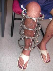
Seeing A Young Boy Wearing An External Fixator and Crutches

Circular External FixatorSometimes I am amazed as what I see when I manage to focus all of my attention on any type of issue related to height, even if the connection is very small. This is what happened yesterday, while I was walking through the famous COEX Mall that is located off of the Samseong Subway Station in Seoul, South Korea. I was having a rough day and I took my GF to the mall to walk around and as we walked around the a mall the sight of a young boy in crutches really caught my eye.

This young boy, who was very young was walking by himself on crutches and had one of his lower legs wearing one of the external fixators which I am so familiar with. The sight was so extraordinary, and when my GF say the metal device that had its metal spoke coming out of his skin, she expressed shock over the fact that this boy was walking around in public which his external fixator. For her, the sight was too disturbing. I felt a little bit of sadness over what this boy was going through. Personally I probably had more of an understanding of why he needed to get this procedure done.

It was clear that this boy was still very young, who was still growing. He was not using the method of distraction osteogenesis to make his legs longer, and look taller. He had a serious medical condition. Most of the procedures of distraction osteogenesis done is to correct for a difference in the length of a certain bone region between the left and right side of a person. You can do callotasis on more than just the long bones, but also the mandibular bone. One of his legs was shorter than the other. To walk with a more normal gait, his leg was being lengthened. I understood that there was a lot of pain associated with the process, but I have never experienced it myself.

This is the first time I have ever seen anyone were an external fixator out in public. However I am not that surprised to see it in a place like Seoul, South Korea. Seoul has been also the same place where I saw a case of a korean women who suffered from gigantism, multiple people suffering from albinism, one case of a person who suffered from Osteogensis Imperfecta (Refer to Sean Stephenson), and various types of achondroplasia and dwarfism. For a country that seems to pride itself on being so homogeneous and image conscious, there are many people who just don’t look ‘average’ whatever that means.

After see the young boy in the crutched, we actually went to donate blood. Apparently blood donating is a popular thing to do these days in Korea. Everyone there in the donation clinic seemed to know what their blood type is, except me. I was once told that my blood type is the most common type there is. The Asian fascination with blood type is something I probably won’t understand very well. It would turn out that my GF’s blood’s plasma is low on Calcium and that her blood did not have the requirements to make her a potential doner. This concerned me greatly, since she has been complaining about how her knees have been hurting for years, the symptom of cold knees, and the desire to be taller. Immediately I realized that there was a clear connection between her lack of calcium, the knee pain, and the cold knees.

How could a women desire to be taller and not even be able to have a sufficient level of Calcium and Vitamin D in her bones and system?

I would immediately take her to the GNC in the mall and tried to stock up on Calcium & Vitamin D 1500 MG as well as Glucosamine Sulphate & Chondroitin 1500 mg. Her resistance on spending $50 on these supplements which would help at least a little with the bone mineral density, combined with her strong desire to become taller, her choice on diet to avoid protein, and her symptom of having intense knee discomfort and cold knees is actually making me realize that she is asking for things in life which are in direct disagreement with each other. She doesn’t know about this website. She doesn’t realize that I have been dedicating for over a year of my life towards this goal, in not just figuring out how to increase height, but also in treating bone and orthopedic disorders. I know how to help her, but she just won’t listen to my advice. Maybe she thinks she is helping me save a little bit of money, but she is risking her own health, and her height by not fixing this problem.

I wrote about the issue of having low Calcium levels leading to severe height loss later in mid age due to menopause in the post The Connection Between Bone Loss From Osteoporosis And Decreases In Height In East Asian Females. The study Usefulness of Estimated Height Loss for Detection of Osteoporosis in Women showed that while a lose of 2-4 cm in height is normal, any more than 4 cms of height loss due to bone density decreases means that osteoporosis has set in. My biggest worry is that not only will she not increase in height, which she desperately desires, due to refusing to listen to my insistence on taking certain supplements to help improve the health of the knees, she is also going to risk getting osteoporosis due to diet choices causing the exact opposite effect for her. 

If she only knew what I know, and saw what I see.