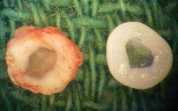
This Researcher Succeeded In 3D-Printing Spinal Discs Allowing Adults With Closed Growth Plates To Grow Taller If They Desired – Big Breakthrough
 My claims and estimates that the technology to find an alternative to limb lengthening surgery would take at least 2 decades, if not 4 decades, may have to be drastically reduced. In my recent post about the nature of how Kurzweil’s Law of Accelerating Return shows an acceleration of technological breakthroughs, it may be that we have already reached the inflection point and things are already accelerating much faster than even I would have anticipated.
The old idea that so many people have suggested of increasing the torso or spine in some way to achieve the goal of increasing their height even as adults with closed growth plates is indeed already here. “How is that possible?” the reader might ask.
The technology has finally arrived, but the biomedical researchers which have succeeded might not even realize just how they can use their new technology in lateral thinking unique and potentially extremely financially rewarding applications.
Update: May 16, 2014: I finally realize now that Dr. Bonassar’s research group had made a very similar ear cartilage tissue like the group from Wake Forest University, which I had referenced twice already on this website. Interestingly he was recently promoted to the rank of Full Professorship back in January of this year, 2014. I guess doing breakthrough research that gets the media and the rest of the world talking definitely helps move one’s academic career much faster. He has quite an impressive Curriculum Vitae, having a BS and MS from MIT in MSE (Material Science & Engineering) and finished his graduate work at JHU (John’s Hopkins University) with his Ph. D in BME (Biomedical Engineering). After getting his Ph.D, he did fellowship work at the Orthopedic Research Lab at Massachusetts General Hospital and then another fellowship at the Center of Biomedical Engineering at his Alma Mater MIT. Before coming to Cornell, he had spent 5 years at the Center for Tissue Engineering at UMass in the Medical School.
So he knows what the hell he is doing and understands tissue engineering and cell culturing and cartilage regeneration much better than me.
I had written quite a few posts talking about how we can use 3D-Printing, and even 4D-Printing of “smart materials” for our endeavor and used his research as an example already. (I also referenced Dr. Valenti’s work on regrowing ear tissue on the sides of lab mice.) The posts were “How 4-D Printing Can Synthesize Biological Tissues And Organs Which Grow Themselves” and “Increase Height And Grow Taller Through Bioprinting And Electrospinning” and “Programmable 3D Printers Creating New Epiphyseal Growth Plate Cartilage As Interchangeable Body Parts“.
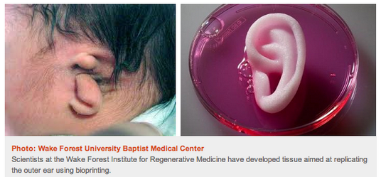 The group from Wake Forest University were the ones who managed to successfully regrow human ears. Remember the picture above??
Even NPR did a short piece on them back in Feb of 2013 on the research going on in Bonassar’s Lab at Cornell (Available Here). Back then, he was still an Associate Professor in BME. Their paper is entitled “High-Fidelity Tissue Engineering of Patient-Specific Auricles for Reconstruction of Pediatric Microtia and Other Auricular Deformities“.
This researcher with his team in some lab at Cornell University has been able to validate and prove a concept which had been another huge block in our progress. You can click below to get the webpage for the Bonassar Research Group, whose goals are focused on regeneration of the musculoskeletal tissue.
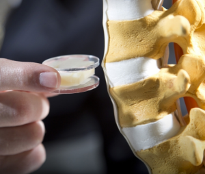
I am referring to the viral post which was written on the website DVice.com entitled “3D printing right into your spine could make you whole again”. I am surprised that neither I or any of the readers of the website ever mentioned it or did not see it written. It proved the validity of an idea which I did not think was reasonable for at least another 10-15 years. However, the whole advent and spread of 3-D Printing technology has made me reconsider so much of what is possible.
So how can we make such a leap in theory from the achievement of regrowing an ENTIRE Spinal Disc, and achieving the goal of increasing our heights as adults?
It turns out that I was wrong before (I admit it. I made a anatomical mistake which led to a bad judgement call, which I will correct right now). Before, I said that there is no way that we should ever touch the spinal disc since we could cause the person in operation to become paralyzed if the spinal cord, which I thought was inside the disc, was nicked or sliced in some way.
Thinking back on it, I realize now that anatomically speaking, it made no sense to assume that the Spinal Cord with the CSF (cerebral spinal fluid) would be in the core of the disc. I was off by around 2 inches at least.
I was reminded of the horrible case of the former Superman actor Christopher Reeves and what he had to go through for over 20 years suffering in a wheelchair after suffering a fall from a horse ride. At the time, I felt that no one should ever be stupid enough to ever choose to go through such an invasive surgery as spinal disc replacement or similar since the risk of paralysis and turning into a paraplegic was just too high, just to gain extra height. If someone did actually go through a surgery like that and risk becoming a paraplegic for just a few centimeters into their torso, they should have their mental state checked out and get major psychiatric help.
Getting back to the question which I still haven’t answered before yet… “So how can we make such a leap in theory from the achievement of regrowing an ENTIRE Spinal Disc, and achieving the goal of increasing our heights as adults?”
So here is what I am going to outline in only semi-technical fashion to show the readers that we already have the technology and the technical know how to achieve this goal.
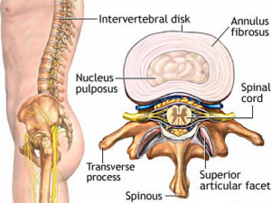 When we actually look at the positioning of the intervertebral disc relative to where the spinal cord is, it seems that they are in different areas of the dorsal region.
The group of researchers was able to using electro-spinning techniques in a Bio-3D Printer to recreate the entire disc, getting the material strength and properties of the nucleus pulposus and annulus fibrosis correct.
Remember that the composition of the NP and the AF are just slight varying percentages of a combination of proteoglycans, collagen, and water.
There is a nice picture that was available on the DVice.com article which I clipped below
This is going to be a 2-Part process which will be combined together in the end for implantation..The steps to do this are the following….
Part 1: For the hyaline cartilage layers…
- Get a sample of the person’s marrow through a bone marrow biopsy. This one is going to hurt quite a bit!
- Take some of the patients own MSCs from their bone marrow through centrifuging and filtration processes.
- Differentiate the MSCs into the chondrogenic linage using growth factory combinations.
- Culture the chondrocytes into cell colonies using a culture medium to increase the cell density
- Take the chondrocytes and using a 3-D Printer layer them into the shape of a thin disc shape. Note that since we are using the 3D Printer to extrude layers upon layer, we can precisely program the alignment of the chondrocytes. With a simple script, we can have the chondrocytes stacked on top of each other like the column-like fashion of the growth plates.
- Don’t forget to also inject the right IHH and PTHrP combination to get the cartilage to expand on its own.
Part 2: For the collagen disc layer…
This is what has been achieved by Bonassar and his research group. They got the structural alignment correct, with the right tissue material properties.
This was one of the other technical problem which I realized we had to figure out, which was shown to have been solved.
Part 3: Re-implantation
- You just layer the cartilage on top and bottom of the collagen disc layer. It turns into a sandwiched multi-layer. This can be implanted into the backs of the patients.
- Since the discs are themselves in the back of the spinal cord, this surgery will be relatively safe from the possibility of cutting the spinal cord.
Part 4: Growth Factor Injections
- After the surgery, the person is recommended to take about 1 week to let the implant to fuse to the vertebrate bones.
- Due to the fact that the printed cartilage sides had the chondrocytes specifically positioned in columnar structures, the cartilage would act like any growth plate and expand in thickness, aka make you grow taller.
- Of course, we recommend increasing the rate of cartilage growth with some carefully positioned growth factor injections, like PTHrP and IHH which I had mentioned before as well as OP-1 (aka BMP-7) which had been proven successful in increasing the disc height of lab rabbits.
- It might even be as simple as using a growth factor patch, where you stick it on your back to let the growth factors diffuse into your back, like the smoking patches you see quitters use.
On The Issue of The Facet Joints Between the Irregular Vertebrate Bones…
- Anatomically, we also have to take into consideration the fact that not only are the irregular vertebrate bones attached to each other through the discs, they are also attached to each other in in the area known as the facet joints, which are areas on the sides where it is ligaments or cartilage.
- I have not done any research on the facet joints, however the joints can be easily be stretched out using injections of a certain type of chemical.
- If the facet joints were mainly ligaments, an injection of the compound relaxin would make the facet joints looser, and easier to stretch out. (The use of relaxin to increase the torso and/or the pelvic girdle bone was the proposed theory me and Tyler gave on why a minority of the female population who went through pregnancy seems to end up taller too.) Even just a few days ago, we had this message to the website where another woman claimed that she increase in height by 2 inches over a span of 2 pregnancies around the age of 33!. (It could be a lie though since we can’t validate all these pregnancy claims, but we wanted to post it here anyway.)
- If the facet joints were mainly elastic or fibrocartilage, a small inject of growth hormone would make the area go into hypertrophy.
The Stretching of the Spinal Cord
This was the other big technical problems we would need to consider. The question is, “If we started to stretch out the torso/back again, would the spinal cord also be stretched out?”
Or is the spinal cord like a piece of string which goes through the vertebrate bone acting like a needle hole?
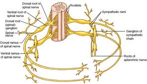 It seems that even a slight bruising of the spinal cord can cause paralysis so we have to be very careful here. It might be that the spinal cord can NOT stretch out or increase in length at the speed of the increasing of the back. If not, the ventral and dorsal roots that laterally comes out of the spinal cord might be pulled out or snapped off, like the branches on a tree.
My personal belief on the elastic nature of the spinal cord says that due to the slow, but noticeable changes in the volumetric size of the irregular vertebrae bones, the spinal cord should not have any difficulty in slowly increasing in small incremental length along with the bone surrounding it.
To all the people who said that Limb Lengthening Surgery is the only way to increase in height, I believe I might have proven you guys wrong, at least in theory. Based on the following steps I have outlined above, there will be some biomedical researcher or company with the funds to take every single step I have outlined above, execute on them, and become a future billionaire.

