Chondrodiatasis Is Tensile Loading The Epiphyseal Growth Plate Cartilage Longitudinally Without Fractures
 For a long time I’ve wondered whether it was possible to do more than just stretch out the bone tissue to make the overall bones longer, and it seems that there have been a few recent articles which came out showing that it is indeed possible to stretch out cartilage (epiphyseal growth plate) tissue. More than just stretching the cartilage, it seems that it would not even result in any type of fractures.
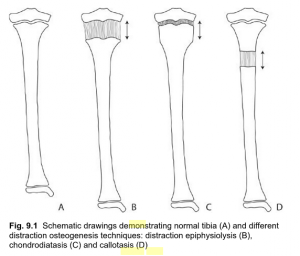
This medical technique is known as chondrodiatasis. It seems that it is so rare and so uncommon that there is almost no information on it except in a few sources from Google. Even orthopedic surgeons are not that too familiar with this idea. (There is a 2nd type of physeal distraction called distraction epiphysiolysis which is much faster but does result in fractures and the cartilage ossifying by the trabecular bone tissue).
In the medical reference book “Musculoskeletal Tissue Regeneration: Biological Materials and Methods“ by Vacanti and Pietrzak there is a small section dedicated to alternative ways to distract bones which is much less step intensive.
There are two types. You have…
1. Distraction Epiphysiolysis – This method is the distraction of the growth plate at a fast rate of 1-1.5 mm per day. The fast rate of separation between the epiphysis and metaphysis results in the cartilage developing fractures, which also leads to the physeal cartilage to turn into trabecular bone. The experiment done by Zavialov and Plaskin back in 1967 showed the change of the cartilage into bone. We won’t focus on this type of physeal distraction technique too much because of the fact that the cartilage is almost immediately converted into bone through advanced levels of osteogenesis.
2. Chondrodiatasis – This is the type of physeal distraction that would be worth our time to look at. It is very slow in fact, usually less than 0.5 mm a day. It seems that tension stress causes the chondrocytes to increase in activity. This technique also increases the level of osteogenesis but there is no evidence (yet) that the cartilage will convert into bone tissue.
Sliedge and Noble back in 1978 showed that they could increase the thickness of lab rabbit’s growth plates by 150% without the presence of fractures. De Bastiani distracted it at just 0.25 mm a day and the chondrocytes cells went through hyperplasia but the overall form of the cells did not seem to change. When the rate of longitudinal growth after physeal distraction was checked, there was no decrease in bone longitudinal growth seen.
Remember however that for any type of bone or cartilage distraction, the surgeons would still drill two pin holes into the bone to pull. It seems that there is no way to get away from having an external fixator used.
However, there will always be complications associated…
Surgical Complication #1: Because we are talking about the growth plates, which are often already so thin, to distract them by only 0.25 mm a day would be very hard to perform.
Surgical Complication #2: The physis would actually get damaged.
Surgical Complication #3: This is the major issue, which seems to be many incidences where growth of the bones seemed to completely stop, which is slightly concerning.
Out of the three ways that you can distract a bone, both of the ways to distract cartilage has proven to be not that effective. That is why the surgeons have almost always just focused on callotasis, instead of these two other types of surgical techniques.
Looking through the PubMed archives, there were probably only a dozen studies which ever mention this term “Chondrodiatasis“. We picked the ones which are worth looking over and linked them below…
- Chondrodiatasis-controlled symmetrical distraction of the epiphyseal plate. Limb lengthening in children.
- Early physeal closure after femoral chondrodiatasis
- A large-deformation, finite-element study of chondrodiatasis in the canine distal femoral epiphyseal plate
We will also make a medical reference to the text “Physeal Injury Other Than Fracture” By Hamlet A. Peterson.
While the initial studies showed us that maybe it is possible to lengthen a bone by distracting the epiphyseal cartilage, which would mean no osteotomy necessary, these later studies showed that maybe it is not such a good idea.
The first study gave us hopes and showed that the surgical technique might be viable. There was no visible lesions, infections, or loss in vascularization.
It is the 2nd study which showed that this technique did have its own share of problems. It seemed that when the bone lengthening was done, almost immediately after the surgery was over, the cartilage ossified over. The end result was that the lengthen bone actually shrank. The end result was that this type of surgery was not recommended for children who are too young or have just a small limb length discrepancy.
The last study done on the femoral growth plates of lab dogs was not as informative as I hoped. All it showed that was if you are going to be pulling the bone, the cortical bone and the area where the cartilage and the bone touch will experience a high level of stress.
Conclusion
These procedures known as Chondrodiatasis and Distraction Epiphysiolysis are not well known because they are probably almost never done anymore. It seems that the amount of complications where the cartilage will ossify to prematurely was the reason most orthopedic surgeons will not try to length bones by doing distraction on the cartilage. Distracting the bone tissue seems to be much less complicated and more straight forward, with much more surgical evidence and examples.

