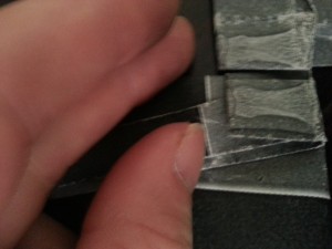
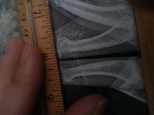
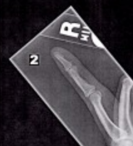
{Tyler’s Notes-Since these measurements are critical to the validity of LSJL I wanted to make a rebuttal right away. Here’s some images that provide evidence that there is in fact a difference between the right and left fingers:
The right finger is at the top. This is the index finger proximal bone. You can very clearly see that the right bone is longer than the left. You can identify that these are the right and left bones on the x-ray page.
Here’s the middle bone:
I apologize for the paper folds confounding where the bone ends and begins but if you look closely you can identify it again. The middle proximal bone is clearly longer. Again the right bone is on the top and the left at the bottom. Michael discusses the protrusion on the bone and suggests it might indicate some type of arthritis and that it might cause bone. What I could do is post a video of myself playing the piano which would provide evidence that my fingers have healthy mobility.
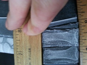
Here’s the proximal part:
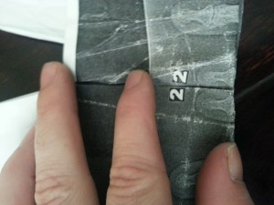 Again right bone is on the top but a length difference isn’t really clear. I’d say this length difference isn’t very significant.
I also did some lateral analysis:
I aligned the bones at the top(proximal) end of the bone. This is the lateral view of the proximal bone of the index finger. Right bone is at the top and left bone is at the bottom. The right proximal index finger bone is longer mainly via the protrusion at the bottom(distal) end. I couldn’t find a significant difference in the middle or top part of the index finger via the lateral view. I also tried comparing the thumb bones and couldn’t find anything noticeable which is odd because my left thumb is now noticeable taller than my right. I’ll have to try to get more accurate measurements there.
How to tell who is right myself or Michael? Print out the x-rays yourself. Cut out each individual bone and compare them against each other.
The main difference and main and Michael’s results is that the left index finger is slanted at about 85 degrees whereas the right index is tilted 75 degrees. Basically, the right index finger is slanting making it shorter. Michael’s theory is that LSJL may have made my finger slanted. However in the X-RAY office the x-ray tech asked me to spread my fingers apart and doing so adjusted the finger tilt.
I know that this proof isn’t optimal(once again) but I wanted to put in some reasonable doubt into Michael’s assertion that my fingers have not grown with LSJL.
If Michael had posted pictures and measurements that shown that I had not grown I would ACCEPT THAT LSJL HAD NOT INCREASED length. Without pictures I can’t analyze his measurement technique. Michael’s conclusion that I have not grown suffers from confirmation bias where he accepts the conclusion that I had not grown from LSJL because that is closest to his beliefs. When I first saw the pictures I too measured that my left index finger was longer until I noticed that the right finger was slanted and had to find a way to adjust this in the measurements.
But I accepted this conclusion because I suffer from confirmation bias that I grew from LSJL. And there’s a definitive and noticeable increase in finger length which is consistent with finger growth(but does not rule out alternative sources). Michael states that he doesn’t have a camera and can’t take pictures of direct measurements. So I’m going to have to do it even if I’m not as skilled(my hands were shaking a little bit when I was trying to take a picture with my camera which made things very difficult).
So hopefull these pictures prove that I noticeably and definitavely now have a right index finger longer than my left and soon I’ll have to step up and take pictures of the measurements.
Read Michael’s analysis below, it is essentially correct you don’t account for that the right finger is more slanted due to the x-ray tech asking me to spread my fingers.}
So in the last week Tyler has sent me a flurry of emails asking that I recheck the blown up X-ray pictures again and measure the entire thing over. Obviously I understand that we need to be extremely accurate on this particular set of X-rays since they would either validate or invalidate his theory LSJL. Here is the set of emails he has sent…
“Did you also do the lateral view for comparison? And did you factor in that the fingers are tilted at different angles? How did you determine the end point of the bone? I would’ve drawn a line at the highest point of each individual bone and then measured between then….Also remember that the right finger is slanted by 10 degree over the left finger.”
“Also you stated you measured the center of the finger which doesn’t take into account the height of the epiphysis of the proximal bone. You can draw a line from the end of that bone and then measure to the end of the finger….And it’s much harder to adjust to the differences in angle for the whole finger if you measure each bone separately it’s easier….To give you an example, I measure the proximal bone of both fingers highest point of epiphysis to lowest point. For left I get 401.0 pixels, in a straight 90 degree line. For right I get 407.0 pixels at a 90 degree angle and the right finger is more slanted….Measure this way and see if you get similar results and we can see if we’re on the same page there at least.”
“Actually what you can do is cut out the bones individually and then it would be easier to align.”
“You stated that you placed the finger bones right next to each other. How did you correct for the 10 degree difference in angle? When you folded how did you align the two papers? I found it was impossible to correct for the angle this way. That’s why I suggested cutting the bones out…Sorry for so many emails but this is obviously very important.”
“Flipping the image may help. I flipped the image with GIMP. Enlarge that image then you can lay them on top of each other and get a better comparison”
“I just did some more measuring. I folded up the papers until only the proximal bones of the fingers were visible. I compared the two bones side to side and used a ruler to align the bones at the bottom. The right proximal bone was noticeable taller than the left. The difference in angle between the two fingers must be partly responsible for the different results. And it seems that each of the three finger bones are aligned a different way so you can’t really compare them accurately side by side because they all slant differently.”
—————————————————-
Here is what I can say after looking over the X-rays this morning. The differences in the lengths, whether I am looking at the most proximal phalanges, or the most distal phalanges all have extremely close/similar lengths, at least for the index finger.
There is a total of 5 pictures, two showing the hands, two lateral pictures of both of the index fingers, and one picture that has the hands side by side.
I have not been able to figure out some type of standard or exact reference point to measure all of these bones by. Bones are irregular shaped and there are no sharp or smooth surfaces to use.
When I tried to measure the epiphysis ends from tip to tip, the measurements came out to be either the same or too close to tell.
Tyler keeps on talking about the fact that the right index finger is at an angle, and I know that. Human bones don’t grow completely straight up.. There will be bound to be a little bit of bending or slanted angle. The question that is probably more important to ask is whether the clamping actually caused the slanting of the right index finger.
This is what I did.
I decided to take the slanted factor out of my analysis. I asked myself just what is going on.
Here is what the X-rays did reveal. It turns out that if you really tried to measure the bones, the most distal phalanges of the left index finger is actually longer than the right one! That would be the opposite of what we are hoping for.
However, the most interesting thing is that the right index finger’s middle phalanges has a completely different epiphysis shape than the left. The distal epiphysis protrudes out twice. That are two pronounced humps on the epiphysis. On the left one, there is just one.
It seems that by clamping that joint for many years, the bone shape of the epiphysis/head has completely changed. It is not smooth with a nice layer of articular cartilage. It is rugged.
That is where the extra length in the finger is from! When I measured the right finger’s middle phalanges, from the most protruded head on the distal epiphysis to the bone’s most proximal edge, it comes out to be substantially longer than if I tried to do it on the left hand. In addition, I did not notice the articular cartilage layer on the epiphysis head in that region. It seems that the clamping might have damaged the articular cartilage layer. In its place is the bulging bone area.
As for the lateral viewpoint of the index fingers, those pictures were the most pronounced. The right index finger had most noticeable thicker bones in the index fingers for the right one. So the bones are indeed thicker. That is where I noticed that the epiphysis head was irregular shaped.
As for the thumbs, I did measure them and could not see any differences that were substantial.
There is a 2nd issue – When I looked at the articular cartilage layer of the most proximal phalanges, it looked like that the right index finger’s one was much thicker than the one on the left. So if you are trying to make the bones thicker, it has noticeable effects.
Conclusion
There are no lateral X-rays pictures of the thumbs to view. The pictures I do have are for the index finger. When measuring the thumbs, I could not find the difference in the lengths of the thumbs.
I conclude now that the reason why the right index finger is longer than the left one is because the clamping has changed the shape of the epiphysis aka the middle phalanges head. The epiphysis is supposed to be thin and mushroom-like. The bone for the right hand has started to bulge out creating two bulges, unlike the one which you see in the X-ray for the left hand. The bulging of the epiphysis on the side is what is elevating the most distal phalanges.
Medical Note: There are 2 things which I should warn people who are potentially going to try the clamping.
Note #1: I don’t see a nice layer of articular cartilage on the distal epiphysis on the middle phalanges of the right index finger. It might indicate that the clamping destroyed the cartilage layer.
Note #2: The articular cartilage for the distal end on the most proximal phalanges of the right index finger looks to be somewhat thicker than the left one. That could mean that the cartilage layer has become inflamed, or maybe it is not.
To conclude the post, I would like to ask Tyler whether he can easily & comfortably bend the tip of his right index finger without pain, since joints without cartilage, aka bone on bone contact, is usually quite uncomfortable, since that is just osteoarthritis. I should know since my grandmother had to have total knee replacement surgery when her knees were just bones rubbing against bones.
For me, even though I have never done the clamping on the index fingers, I can’t bend the most tip part of my right finger very well. It would seem that maybe Tyler hasn’t noticed that it has become maybe progressively more uncomfortable or painful to bend that right index finger tip.
LSJL seems to have proven that it can make bones thicker, and can even change the head of long bones aka epiphysis into different shaped. Technically, LSJL does increase the length of the bones, when measured from the end points tip to tip, but it does the lengthening by altering the epiphysis to a shape which is not really normal. The surface of the top of the epiphysis is rugged/no longer smooth. The articular cartilage seems to become thicker, and become ossified. Whatever is below that joint obviously gets pushed in the distal direction. If we are talking about clamping the knees, the tibia/fibula/calcaneus/other feet bones will all be pushed downward. Technically the net result is an increase in height.
Many people who have been critics of the LSJL theory (like those on the Sceptic Forums, available here) claim that the clamping will cause the cartilage to swell up and lead to osteoarthritis later on. At this point, the X-Rays seem to validate their concerns.

