Tag Archives: LSJL

LSJL research lives. A student references lateral loading in a paper

I was worried about the future of Lateral Synovial Joint Loading with most of Hiroki Yokota’s new papers being focused on other things but it seems like a researcher by the name of Zengphei Zhang has spoken a lot about the lateral loading technique in a new paper. I don’t know if Zengphei Zhang is related to Ping Zhang who also worked on Lateral Synovial Joint Loading. But that would explain his emphasis on it. However, regardless it is exciting that research is being done in the field. Zengphei Zhang has used a lateral loading device before in this study. He references this manuscript “Mechanical Stimulation: A Non-invasive Treatment Strategy for Leg Length Discrepancy” that might have additional info but I can’t find it.

Effects of mechanical load/stress on bone growth

“Leg length discrepancy (LLD) is a condition characterised by a difference in bone length, where one leg is shorter than the other. Surgical treatments for LLD are often associated with complications such as pain at the surgical site, infection, and delayed bone union. Thus, there is an ultimate need to establish noninvasive approaches to treat LLD. This thesis explores the use of mechanical loading as a potential noninvasive method to treat LLD.”<-It’s very exciting that mechanical loading is being used as a potential method to treat limb lengthening discrepencies. Even if they suspect that mechanical loading works via the growth plate that does not mean that a non-growth plate mediated mechanism does not exist. There are cases of bone growth increasing in length past puberty. Also there is a study which shows that lateral loading can increase longitudinal bone growth in growth plates that are near senescent. Thus, lateral loading could potential reverse growth plate senescence and could potentially awaken growth plates.

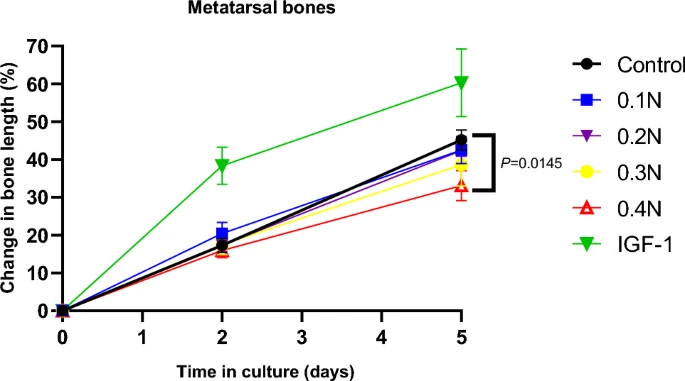
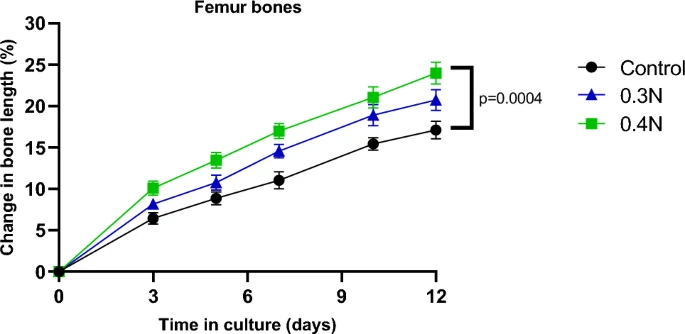
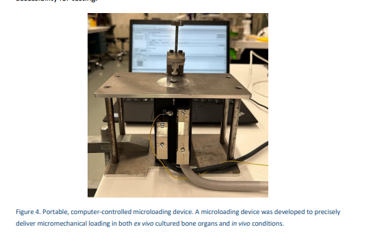
“I focused on the creation of a portable, computer-controlled microloading device capable of delivering precise mechanical loading to small bone organs and animals, such as mice. Using this device, we tested the direct effects of mechanical forces on rat embryonic femur and metatarsal bones cultured ex vivo. These results revealed that mechanical loading at 0.4N significantly decreased growth in metatarsal bones (by approximately 1 mm) while significantly increasing growth in femurs (by approximately 4 mm). These findings suggest that the impact of mechanical forces on bone growth appears to be influenced by both the size and unique traits of the bones.” Maybe the device is too large for the smaller metatarsal bones. In which case just making a smaller device would work. It’s possible that loading at a relatively smaller surface area is optimal.

“Study III extended these findings to young mice, where mechanical loading was applied to the joints of one hindlimb (both female and male, 4-week-old and 8-week-old){unfotunately, these mice are pretty young}. Mechanical loading significantly increased femur length, with the most pronounced effects observed in 4-week-old mice of both sexes. Furthermore, we identified PTGS2 (prostaglandin- endoperoxide synthase 2) as a key gene involved in the bone-lengthening effects of mechanical loading. PTGS2 expression was significantly elevated in the CD73+ and PTHrP+ skeletal stem cell niches of the growth plate in treated legs. Pharmacological inhibition of PTGS2 abolished the bone-lengthening effect, confirming its critical role. Furthermore, mechanical loading significantly increased both PTGS2 expression and the size of ex vivo cultured human growth plate cartilage.”

Soft tissue constraints being mentioned also indicates that we could find some way to reduce the soft tissue constraints in order to grow taller.

“the parameters of mechanical load, including load regime (static or dynamic), force level, frequency, orientation (axial or lateral), and age and growth plate direction, are not taken into consideration. “

“Limited studies report the effect of dynamic lateral load on longitudinal bone growth. Zhang’s group, using a customised piezoelectric mechanical loader, applied a sinusoidal force laterally to the mouse knee joint and examined the length of the hind limb two weeks after the last loading. The force was given precisely at 0.5N with 5Hz for 3 min/day, 10 days in total. It was found that dynamic lateral loading increased the femur length by 3.5% and 2.3% when compared to the age-matched control and contralateral non-loaded side, respectively. It was the first study showing that laterally applied dynamic load may enhance longitudinal bone growth. However, there are several limitations of this study, including the absence of data on any gender- or age-specific differences, as well as insufficient mechanistic studies. Similarly, in 8-week-old female mice, the same group investigated the effects of mechanical force and frequency administered for 5 min per day laterally to the elbow joint for 10 days.

They observed that in comparison to the contralateral non-loaded side control and age-matched control, the length of humerus was increased by 1.2%. While a greater increase in ulna length was also observed, with 1.7% and 3.4% growth compared to contralateral control and age-matched control, respectively. Notably, these effects were accompanied by increases in body weight along with bone mineral density and content.”<-this is a direct reference to lateral compressive load to the epiphysis of bone.

One study did find “dynamic axial loading at 5 N of 2 Hz significantly enhanced longitudinal bone growth of tibia bone in mice” but there were other studies that showed growth inhibition due to axial loading.

“The very common belief is that certain sports can alter an individual’s final stature. For example, weightlifting is thought to decrease body height, whereas basketball is believed to increase it. This is probably due to the individual’s biotype, which means tall individuals are more likely to choose to play basketball, while short individuals like to participate in weightlifting. Indeed, a study in gymnasts showed that short stature and delayed puberty are due to selection bias on training period and leg length. Similarly, another study reported that the short stature found in gymnasts is due to selection bias”<-this is the first paper I’ve read that mentions the height basketball correlation.

“Static compressive mechanical load has been reported to diminish the type II collagen (Col 2) expression and type X collagen (Col X) expression in the hypertrophic zone of tibial growth plates in rat”

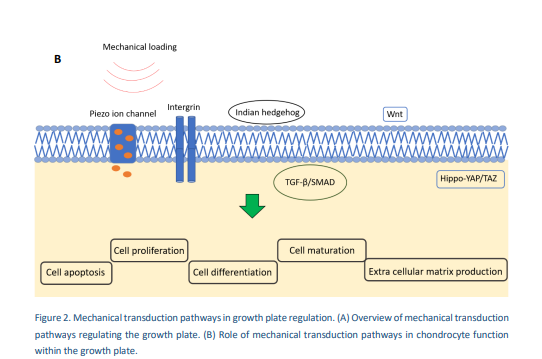
“Mechanical load plays a vital role in regulating chondrocyte activity within the growth plate, with Piezo ion channels (Piezo1 and Piezo2) are important mechano-transducers which convert mechanical force into electrochemical signals. Notably, Piezo1 and Piezo2 have been found largely to be expressed in the chondrocytes, and it was observed that mechanical stress can cause Ca2+ to flow into chondrocytes through Piezo channels, resulting in cellular apoptosis. Interestingly, the endogenous peptide urocortin, expressed in primary human articular chondrocytes, has been found to block the Piezo1 channel in vitro, which ultimately protects chondrocytes from apoptosis. Further, inactivating Piezo1 in chondrocytes drives a disturbance in endochondral bone formation, which is highly regulated by the growth plate”

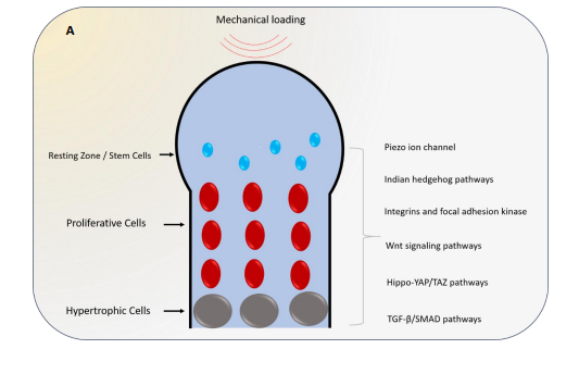
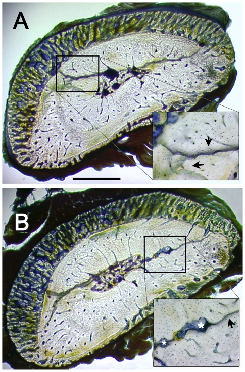
The above shows how loading could alter the growth plate. Note that if mechanical loading could impact stem cells it could potentially form new growth plates. Another image of how mechanical loading could modulate longitudinal bone growth:

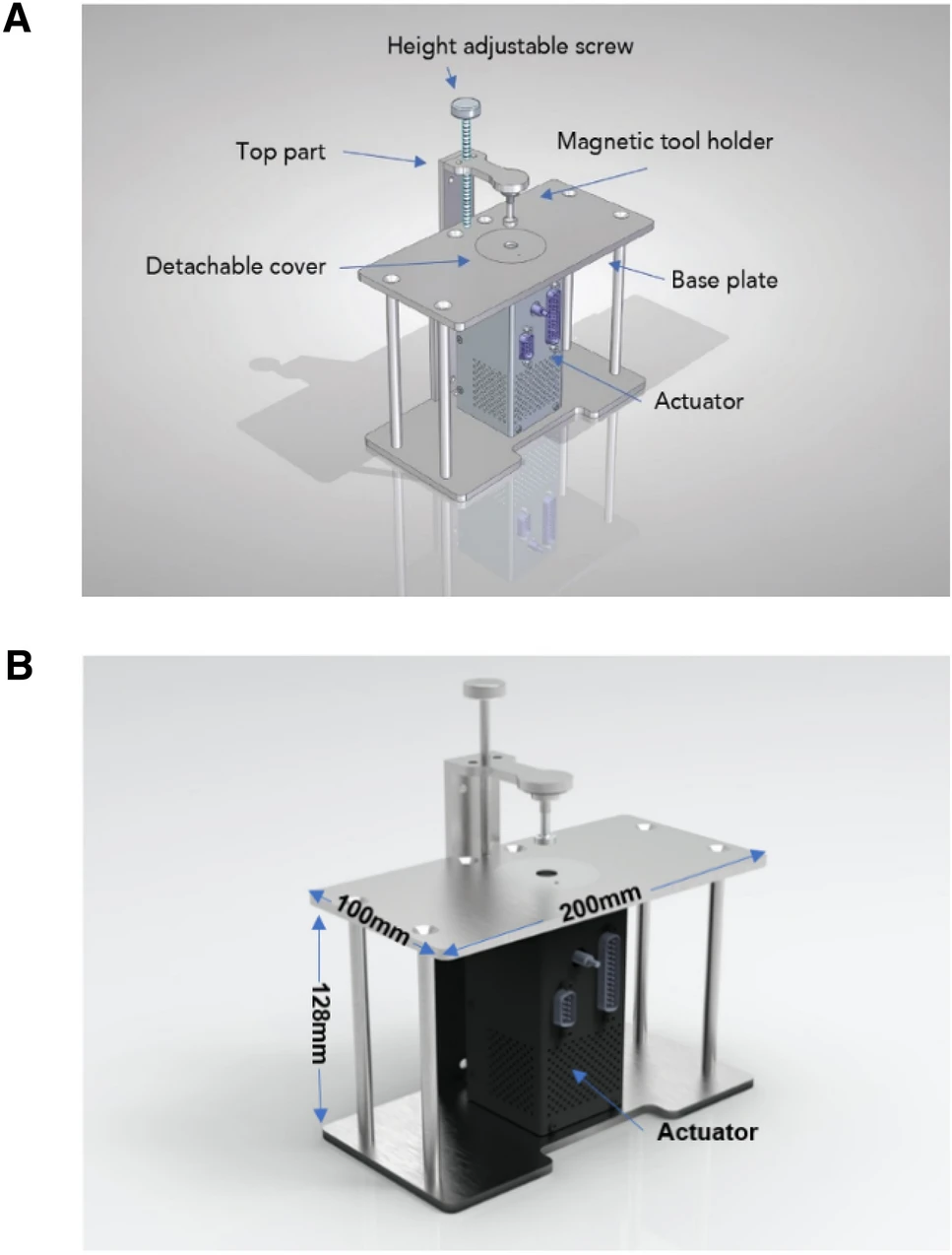
Here’s a picutre of the device, you can see why it may be too large for smaller bones:

“Mechanical loading significantly enhanced femur length growth in loaded hindlimbs compared to sham-loaded hindlimbs in all mice, regardless of age or sex. The increase in length was consistent at both the 14-day and 28-day time points, with the more prominent effects observed when the load was applied daily rather than every other day.”

“Mechanical loading significantly increased height of the growth plate in the loaded hindlimbs at both 14 and 28 days. A higher number of proliferative and total cells in the growth plate regions with notable differences were also observed at both time points. Additionally, the hypertrophic zone height was significantly greater in loaded limbs, while the size of individual hypertrophic chondrocytes remained unaffected.“<-perhaps lateral loading can also increase the height of the articular cartilage contributing to height that way.

Mechanical loading of human growth plate cartilage ex vivo resulted in significant increases in growth plate height and cartilage growth after 28 days. The PTGS2 and PIEZO1-positive cell numbers were significantly higher in loaded samples compared to sham-loaded controls, confirming that the mechanisms observed in mice also apply to human growth plate tissues.”<-a common criticism of LSJL is that it has only been shown to work in rats and not humans but this indicates that this scientist believes that lateral loading could work in humans. But this does not yet indicate that Lateral Dynamic Loading will work on adults post skeletal maturity.

Huge news someone other than Yokota/Zhang use a joint loading device

This is huge that someone else is doing a LSJL like device it means that something could be close to being put into practice.

Micromechanical Loading Studies in Ex Vivo Cultured Embryonic Rat Bones Enabled by a Newly Developed Portable Loading Device

Mechanical loading has been described as having the potential to affect bone growth{we want it to affect bone growth post skeletal maturity of couse}. In order to experimentally study the potential clinical applications of mechanical loading as a novel treatment to locally modulate bone growth, there is a need to develop a portable mechanical loading device enabling studies in small bones. Existing devices are bulky and challenging to transfer within and between laboratories and animal facilities, and they do not offer user-friendly mechanical testing across both ex vivo cultured small bones and in vivo animal models. To address this, we developed a portable loading device comprised of a linear actuator fixed within a stainless-steel frame equipped with suitable structures and interfaces. The actuator, along with the supplied control system, can achieve high-precision force control within the desired force and frequency range, allowing various load application scenarios{the potential would be to use the device to induce longitudinal bone growth on skeletally mature individuals}. To validate the functionality of this new device, proof-of-concept studies were performed in ex vivo cultured rat bones of varying sizes. First, very small fetal metatarsal bones were microdissected and exposed to 0.4 N loading applied at 0.77 Hz for 30 s. When bone lengths were measured after 5 days in culture, loaded bones had grown less than unloaded controls (p < 0.05). Next, fetal rat femur bones were periodically exposed to 0.4 N loading at 0.77 Hz while being cultured ex vivo for 12 days. Interestingly, this loading regimen had the opposite effect on bone growth, i.e., loaded femur bones grew significantly more than unloaded controls{femurs have different proportions than metarsal bones, one possibility is that femur bones shape makes it more susceptible to fluid based forces and pressure gradients, due to the femurs longer shape it is possibly more susceptible to deforming forces than metatarsals}. These findings suggest that complex relationships between longitudinal bone growth and mechanical loading can be determined using this device. We conclude that our new portable mechanical loading device allows experimental studies in small bones of varying sizes, which may facilitate further preclinical studies exploring the potential clinical applications of mechanical loading.”

“two use-cases were devised: repetitive impact loading of small force (0.05–0.5 N) at a range of  30–180 repetitions and continuous sinusoidal loading of medium force (0.5–5 N) at a frequency range of 5–20 Hz.”<-something like a massage gun does something similar of repetitive impact loading.

Above is the device used.

So unfortunately for metarsals it does look like yes the device suppressed growth. However, initially the 0.1N load enhanced growth. More studies would have to be done.
In contrast femur bones grew pretty uniformly and the difference is pretty significant. Again, I think it is possible that this could be due to the different shape of the femur bones or maybe the load was too strong for the smaller metatarsal bones.

“Its square shape securely covers the entire cartilage area on each side of the embryonic femur bone, allowing the indenter to apply mechanical loading specifically to the growth plate in a stable manner.”<-just because the growth plate was the thing that was loaded does not necessarily mean that the growth plate was solely responsible for the growth there could be effects elsewhere as well including that which induces longitudinal bone growth.

“we identified that the same load applied to bones of different dimensions has opposing effects on bone growth, suggesting that the effects of mechanical loading on growth are dependent on the magnitudes and relative dimensions of the bones.”<-so they too think that the shape of the bone has an impact on whether growth is induced or not. The fact that the shape of the bones matter also suggests that yes it is possible that not all of the growth is due to the growth plate.

“Embryonic metatarsal bones from Day 19.5 of gestation are approximately 1 mm in length, whereas femur bones are approximately 4 mm, indicating that the level of mechanical loading that stimulates or inhibits bone growth is likely dependent on bone size.”

This is the sentence where the cite Yokota/Zhang’s joint loading study: ” The device’s controller enables sinusoidal loading, which has been demonstrated to have a bone growth-promoting effect in mice “

Can Flexioss be used to to prove LSJL or Lateral Impact Loading?

Arthur Lazar is someone who has spoken about LSJL in the past on Quora. “Not really. There is 0 evidence for that. The original working experiment was performed on mice – mice growth plates never ossify. MAYBE if someone would develop a machine which can put perfect constant pressure, perfectly shaped for bone area where the pressure is supposed to be, then in theory it could work. But this is a bro-science, so it’s a big MAYBE. But as for now, using clamp, dumbelss or whatever you can use to press at bone would never work.”<-Mice growth plates don’t ossify but they become senescent which is just as bad for growth.

Here’s some more of his thoughts on LSJL: “Yes, I do work on a device for automatic long bone loading method as I believe that the standard lsjl loading (manual with clamps, weights, mpistols) is an invalid approach that lacks consistency, frequency and stability which all was provided with the original, successful experiment.”<-I don’t know what an mpistol is. I believe it is a typo. I don’t know what the original intent is.

“Thank you for your interest, but currently my team is complete and current priority of the projects puts the lsjl idea on the bottom of the list. When I am done with the prototype and IF it will have a desired affect on Flexioss structure (in the terms of force application on the structure) I will publish the design in order to expand the team and get potential investors interested.”

Here’s another set of communications someone had with him.

So the question is should we be using flexioss to try to find the best loading regime to induce the proper stimulation to induce new longitudinal bone growth. I believe personally that the best regime is some kind of lateral impact loading(I believe that tapping the epiphysis would be superior than the diaphysis now but I am trying both). Clamping has a slippage problem which impact does not have. The loads of direct lateral impact are stronger than that occur during normal physiological activities which are more axial.

Lateral impact does occur during boxing both to the hand and to the face and ribcage. Also, it occurs to the feet bones during running(but this depends on whether you are a heel or toe striker). It also happens to bones during muay thai kick boxing.

The problem is that this impact is often at irregular intervals and not targeted to specific areas of the bone such as the epiphysis. The epiphysis is where there is less cortical bone, is close to where the growth plates used to reside in skeletally mature individuals, and is close to the articular cartilage which if stimulated could potentially contribute to height growth. In muay thai you have no control over where you are kicked and if you do kick you are trying to use the strongest part of the bone.

Lateral impact has the potential in my opinion to drive the most fluid forces throughout the bone. Greater than any axial impact certainly due to the pressure gradient of the bone and the epiphysis is the weakest most porous part of the bone so impact to that area has the potential to drive fluid forces throughout the entire bone. Muscular contractions also have the ability to stimulate fluid forces throughout the bone but that is limited by muscular size and strength. Lateral impact also has the ability to gradually induce plastic deformation throughout the bone. Most plastic deformation occurs axially to shorten the bones such as in rickets/paget’s disease etc. Lateral impact loads have the potential to induce plastic deformation in a way such as to lengthen the bones.

Here is the flexioss.

So the question is can we use the flexioss to find the best way to induce lateral plastic deformation in such a way as to lengthen the bones or to induce fluid forces to either induce articular cartilage endochondral ossification or to cause denovo cartilagenous regions within the bone.

In the study Dose-dependent new bone formation by extracorporeal shock wave application on the intact femur of rabbits., they found trabecular bones heaving with cartilagenous tissue which would be huge as bone tissue is not capable of interstitial bone growth.

The manufacturers of flexioss claim that it has properties similar to that of cancellous bone so yes it can potentially be used to find the best loading regime to induce plastic deformation in such a way as to longitudinally lengthen the bone. Obviously, it can’t really be used to mimic the fluid properties of the bone.

Microcracks effect on fluid flow. Long term loading may be key for LSJL.

This study is important because it indicates that microcracks may be bad for fluid flow which stimulates bone growth but good for cartilage growth as if fluid is not flowing than it is building up pressure and pressure is more conducive to chondrogenesis.  This indicates that a method to induce fluid flow such as  clamping/tapping should be higher duration and more “fatigue loading” based to insure the induction of microcracks.  Fatigue loading is the act of inducing bone damage not by a sudden large damage but by sustained bouts of loading over time.

Influence of interstitial bone microcracks on strain-induced fluid flow.

“microcracks act as a stimulus for bone remodelling, initiating resorption by osteoclasts and new bone formation by osteoblasts.  Microcracks alter the fluid flow and convective transport through the bone tissue. [We evaluate] the strain-induced interstitial fluid velocities developing in osteons in presence of a microcrack in the interstitial bone tissue. Based on Biot theory in the low-frequency range, a poroelastic model is carried out to study the hydro-mechanical behaviour of cracked osteonal tissue. the presence of a microcrack in the interstitial osteonal tissue may drastically reduce the fluid velocity inside the neighbouring osteons{So maybe microcracks will increase hydrostatic pressure as hydrostatic pressure is the pressure exterted by a fluid at rest}. This fluid inactive zone inside osteons can cover up to 10% of their surface. Consequently, the fluid environment of bone mechano-sensitive cells is locally modified.”

“Cortical bone constitutes the outer shell of long bones. This live entity is continuously renewed by bone cells in response due to the loading generated by daily activity”

“microdamage occurring inside the osteonal volume may generate a cell-transducing mechanism based on ruptured osteocyte processes. Concomitantly, microcracks are likely to alter the fluid flow and convective transport through the bone tissue and thus modify the hydraulic vicinity of the sensitive cells”

“the drag force caused by the pericellular fibres is thought to activate the cellular biochemical response through the interactions with the cytoskeleton”

“the pressure inversely increases from its Haversian reference to reach its maximum in the interstitial tissues.”

“the presence of the microcrack strongly modifies the fluid flow velocities in the osteons located in the immediate vicinity of the damage. It may generate an “inactive zone” inside the osteon wherein the fluid velocities are relatively low and thus the osteocytes stimulation too”<-But even though fluid flow may be low hydrostatic pressure may be high which may be better for chondrogenesis.

Fatigue Loading may be important to LSJL

This paper shows that axial loading can induce an almost complete fracture line through the bone through one side to the other.  But the break is on the wrong axis.  If fatigue loading was induced via transverse loading (lsjl or lateral impact loading) it is very likely that the micorodamage would be along the right axis.  But to induce bone fatigue in this way would likely require heavier loads than inducing sufficient hydrostatic pressure to induce chondrogenesis as the goal of increasing fluid forces in the bone is to encourage bone decay and induce differentiation of tissues that are capable of interstitial growth.

Role of Calcitonin Gene-Related Peptide in Bone Repair after Cyclic Fatigue Loading

“We used the rat ulna end-loading model to induce fatigue damage in the ulna unilaterally during cyclic loading. We postulated that CGRP would influence skeletal responses to cyclic fatigue loading. Rats were fatigue loaded and groups of rats were infused systemically with 0.9% saline, CGRP, or the receptor antagonist, CGRP8–37, for a 10 day study period. Ten days after fatigue loading, bone and serum CGRP concentrations, serum tartrate-resistant acid phosphatase 5b (TRAP5b) concentrations, and fatigue-induced skeletal responses were quantified. cyclic fatigue loading led to increased CGRP concentrations in both loaded and contralateral ulnae. Administration of CGRP8–37 was associated with increased targeted remodeling in the fatigue-loaded ulna. Administration of CGRP or CGRP8–37 both increased reparative bone formation over the study period. Plasma concentration of TRAP5b was not significantly influenced by either CGRP or CGRP8–37 administration.”

“sensory innervation of bone may have regulatory effects on skeletal responses to bone loading”

“Periosteum, endosteum, and bone tissue are all innervated by nerve fibers. This innervation exhibits plasticity in response to mechanical loading, in that a single loading event results in persistent changes in neuropeptide concentrations in both loaded and distant long bones, as well as changes in the neural circuits between limbs

Individual bone cells are directly connected to the nervous system via unmyelinated sensory neurons. Bone cells express a range of functional neurotransmitter receptors and transporters, including those for calcitonin gene related peptide (CGRP)”

“12 rats were fatigue loaded until 40% loss of stiffness was attained, using an initial peak strain of −3,000 µε (Fatigue group). ”

“To induce fatigue, the load applied to the ulna was incrementally increased until fatigue was initiated, as indicated by increasing displacement amplitude from a stable baseline. ”

The break extends all along the bone but is on the wrong axis.
“Increased bone blood flow precedes bone repair in response to fatigue loading, and remodeling in response to decreased mechanical loading . Systemic administration of CGRP also decreases blood pressure in a dose-dependent manner””
“”treatment with CGRP8–37 may have increased intraosseus pressure, transcortical interstitial fluid flow, and associated bone formation””

New study shows LSJL induces Bone Deformation

Bone Deformation is change in the bone shape or structure.  This deformation can be compression of various cavities, stretching of the bone, twisting, and so son.  This is important as bone deformation is one way to increase hydrostatic pressure by decreasing the cavity size.  Hydrostatic pressure is the pressure exerted by a fluid at rest.   Compressing the bone laterally inhibits the fluids ability to move thus increasing hydrostatic pressure.   If there is more fluid within a smaller space than it follows that hydrostatic pressure increases. Hydrostatic pressure has been consistently shown to induce chondrogenic differentiation.  Chondrogenic tissue is the key for longitudinal bone growth as traditionally chondrogenic tissue is capable of interstitial(growth from within) whereas bone is not.  Only interstitial bone growth has been shown traditionally to induce significant longitudinal bone growth but there are potentially other ways to stimulate longitudinal bone growth.

Knee loading inhibits osteoclast lineage in a mouse model of osteoarthritis.

“Osteoarthritis (OA) is a whole joint disorder that involves cartilage degradation and periarticular bone response. Changes of cartilage and subchondral bone are associated with development and activity of osteoclasts from subchondral bone{Since osteoarthritis does affect the subchondral bone that does affect our ability to say that LSJL affects bone deformation in a normal bone but there is no reason why it shouldn’t}. Knee loading promotes bone formation. Knee loading regulates subchondral bone remodeling by suppressing osteoclast development, and prevents degradation of cartilage through crosstalk of bone-cartilage in osteoarthritic mice{This “crosstalk” may stimulate chondral tissue within the bone as well}. Surgery-induced mouse model of OA was used. Two weeks application of daily dynamic knee loading significantly reduced OARSI scores and CC/TAC (calcified cartilage to total articular cartilage), but increased SBP (subchondral bone plate) and B.Ar/T.Ar (trabecular bone area to total tissue area). Bone resorption of osteoclasts from subchondral bone and the differentiation of osteoclasts from bone marrow-derived cells were completely suppressed by knee loading{Knee loading affects the differentiation of bone marrow-derived cells which is the first step in proving that it causes chondrogenic differentiation}. The osteoclast activity was positively correlated with OARSI scores and negatively correlated with SBP and B.Ar/T.Ar. Furthermore, knee loading exerted protective effects by suppressing osteoclastogenesis through Wnt signaling. Overall, osteoclast lineage is the hyper responsiveness of knee loading in osteoarthritic mice. Mechanical stimulation prevents OA-induced cartilage degeneration through crosstalk with subchondral bone. Knee loading might be a new potential therapy for osteoarthritis patients.”

“Daily dynamic knee loading was applied at 1 N, 5 Hz, 5 min/day for 2 weeks”

joint loading on subchondral bone

Compare the OA+ loading to the control bone.  The subchondral bone plate looks much more dynamic.  There are three bone marrow regions rather than two(bone marrow is the blue dots).

You’ll also note that loading+OA increased the ratio of calcified cartilage out of total articular cartilage(but not above statistic significance.  It did not fully restore the thickness of the bone plate.  Alendronate is an anti reobsorption agent.  Given that the ALN and loading group is different we can say that change in subchondral bone shape is likely not related to inhibiting osteoclast activity and is something unique to the loading group.

loading affect on subchondral bone2

Compare the joint capsule region of control and OA+loading group.  The Joint Capsule is the region that’s not inside the bone.  The cells are a lot more spread out.  There’s a dense redness in the control group which is not present in the OA+Loading group

We can see that the loaded group again has distinct characteristics and we can also see the growth plate.  The growth plate of the LSJL group is distinct and there does seem to be signs of cellular migration.  I’ll have to blow it up.

cell migration

Circled is the region of possible cell migration.

LSJL-growth-plates

Similar signs of migration in earlier in LSJL studies(above taken from Lengthening of Mouse Hindlimbs with Joint Loading).

“the expression of Wnt3a was significantly increased by knee loading. However, the protein and mRNA levels of NFATc1, RANKL, TNF-α, and Cathepsin K were significantly suppressed by knee loading”

“Female C57BL/6 mice (~14 weeks of age)”

If you look at this image of bone marrow derived cells extracted from the loaded group and the other groups you can see that the cells are more condensed and condensation is a prerequisite for chondrogenic differentiation,

Loaded cells are more condensed

This is an image of what mesenchymal condensation looks like:

mesenchymal condensation