Tag Archives: LSJL studies

LSJL Studies 4: Some LSJL effects summarized

Unfortunately, not a lot of insight for LSJL for height growth as most of the effects are those related to the ability of joint loading to inhibit catabolism.

Mechanical intervention for maintenance of cartilage and bone.

Moderate loads to the synovial joint suppress the expression levels of matrix metallproteinases (MMPs), while loads above a threshold tend to increase their destructive activities{although some catabolic effects of MMPs may be good for height growth, as MMPs may degrade bone allowing for cartilage growth}.

“Moderate shear stress(2–5 dyn/cm2) reduced MMP expression levels, while high shear stress (10–20 dyn/cm2) increased them. Moderate hydrostatic pressure (1–5 MPa) suppressed MMP-1 expression, while higher loads (10 MPa) elevated it.”<-Since I have gotten more results with higher clamping force it could indicate that increased MMP expression is crucial to induce new length growth.

“The required magnitude of loads for joint loading is in general smaller than that for axial loading (e.g. 0.5 N for elbow loading and 2–3 N for ulna axial loading in mice). Bone is less stiff in a lateral direction than an axial direction.”<-Note that more than 0.5N(100N is mentioned) is likely required for humans. 0.5N is what was used in the mouse arm lengthening study.

“Joint loading periodically alters the pressure in the medullary cavity and activates molecular transport in a lacunocanalicular network in cortical bone.”<-It is our hypothesis that this increase in pressure in the medullary cavity induces chondrogenic differentiation.  The medullary cavity is continuous into the spaces of the spongy bone of the epiphysis.  It is these spaces where we aim to induce chondrogenic differentiation and thus induce endochondral ossification to grow taller.

“A pressure gradient in the medullary cavity generates oscillatory fluid flow in the porous bone cortex. Induced fluid flow then enhances molecular transport in the lacunocanalicular network and applies shear stress to osteocytes residing in lacunae”

“A pressure gradient in the medullary cavity generates oscillatory fluid flow in the porous bone cortex.”<-and fluid flow into the spongy bone spaces of the epiphysis and the increased MMP expression could allow the neo-growth plates to spread to other parts of the bone but this is highly speculative.

“Modulation of the intramedullary pressure with knee loading is exerted throughout the length of the tibia and the femur.”<-the epiphysis is part of the entire length thus knee loading like by LSJL alters pressure in the epiphysis.

“Proinflammatory cytokines such as IL-1β upregulate the expression and activity of MMP-1 and MMP-13. [In] cultured chondrocytes mechanical stimulation, given in a form of fluid flow shear stress, can suppress the IL-1β-induced upregulation of MMP-1 and MMP-13. In accordance with those in vitro results, joint motion in vivo is able to reduce inflammatory responses in a murine collagen-induced arthritis model. Additionally, in an antigen-induced arthritis model in rabbits, continuous passive motion suppressed transcription of IL-1β and synthesis of inflammatory mediator COX-2 and MMP-1. These mechanical signals also induced IL-10 synthesis, suggesting that moderate joint loading can generate anti-inflammatory signals.”<-MMP’s have complicated effects on height growth.  MMP-1 and MMP-13 are vital for the endochondral ossificatiion process.

“When knee loading was applied to one leg, the loaded tibia and femur were reported to be longer than the non-loaded contralateral bones. In response to knee loading, the number of cells in the growth plate of the proximal tibia increased and their cellular shape was altered.“<-If LSJL increases the number of cells in the growth plate by differentiation of stem cells into chondrocytes than LSJL will work in adults as well.  It’s possible that during knee loading only chondrocyte proliferation was increased but chondrocytes have a finite proliferative capacity and an increase in chondrocyte proliferation without increasing stem cell differentiation into chondrocytes should accelerate the the transition of proliferating chondrocytes into hypertrophic chondrocytes and not the number of cells in the growth plate.

Homeostasis of the articular cartilage is affected through interactions with the subchondral bone underneath the cartilage.  Both MMPs and ADAMTS need to be post-translationally activated, and this activation process is regulated by many factors including MMPs themselves and many proteoglycans.”<-Thus loading of the articular cartilage may itself play a role in the height gain by triggering a response in the subchondral bone in response to the stimulation of the articular cartilage.  Thus underlying the importance of loading the synovial joint.  The activation of MMPS and ADAMTS in the subchondral bone may play a role in neo growth plate formation.

“flexion of the joint in the presence of axial loads (5 N) increased the level of MMP-13 mRNA and its activity.”<-LSJL height growth can not be due to higher levels of MMP13 alone or axial loading would increase height!

“Whether mechanical loading can suppress or induce the integrated stress response is largely dependent on the loading intensity. This stress response leads to translational de-activation by a mechanism involving phosphorylation of eIF2α, with preferential translational activation of a particular set of proteins linked to cellular survival or apoptosis. In cultured chondrocytes, administration of thapsigargin and tunicamycin induces stress to the endoplasmic reticulum, which triggers an integrated stress response. In this response, the level of phosphorylated eIF2α was elevated together with the expression of MMP-13. Joint loading reduced the level of phosphorylated eIF2α by suppressing activity of Perk, one of the four known eIF2α kinases”

LSJL Studies 3: Lengthening of mouse hindlimbs with joint loading

This is the most significant LSJL study to date.

Three key takeaways from this study:

1) LSJL increases bone length in existing growth plates via traditional mechanisms(chondrocyte hypertrophy)

2) LSJL increases bone length in non-traditional mechanisms as shown by the fact that LSJL also stimulates the reserve zone.  Reserve zone cells being the chondrocyte precursor cells and the ones able to form new growth plates.

3) LSJL dramatically alters the microenvironment of the bone(as shown by the histological slides).  It’s unclear exactly what changed but the decrease in bone trabeculae and the increase in bone marrow means that an LSJL loaded bone is more permissive to growth plate formation.  Osteomy is essential for renewed longitudinal bone growth.  As cartilage is capable of interstitial growth which induces longitudinal bone growth whereas bone is not.

Lengthening of mouse hindlimbs with joint loading

“Loads were applied to the left hindlimb (5-min bouts at 0.5 N[at 5Hz) of C57/BL/6 mice (21 mice, ~8 weeks old). Compared to the contralateral and age-matched control groups, knee loading increased the length of the femur by 2.3 and 3.5%, together with the tibia by 2.3 and 3.7%, respectively. In accordance with the length measurements, knee loading elevated BMD and BMC in both the femur and the tibia. Histological analysis of the proximal tibia revealed that the loaded growth plate elevated its height by 19.5% and the cross-sectional area by 30.7%. Particularly in the hypertrophic zone, knee loading increased the number of chondrocytes as well as their cellular height along the length of the tibia.”

3min/day for 5 days/week for 10 days total was LSJL applied.  Bone was harvested 18 days after the last loading.

“Femoral length was defined as the maximum distance from the distolateral condyle to the
most medial and proximal position on the femoral head. Tibial length was defined from the most proximal position of the tibial plateau to the most distal position of the medial malleolus.”<-this is important as changing where and how femoral length is measured would effect total femur length.  It is hard to tell the ramifications of this length setting for sure without more data though.

“The height of the growth plate (GP) was defined from the apical[apex] border of the reserve zone to the lower border of the mineralized cartilage”<-So the measurement of growth plate height would likely include not just growth plate chondrocytes but chondrocyte progenitor cells.  And you’d need chondrocyte progenitor cells to form new growth plates.

According to Artificial selection sheds light on developmental mechanisms of limb elongation, an increased number of proliferative chondrocytes is likely the cause of increased height.

“the upper boundary of the hypertrophic zone was identified at the margin of the
chondrocytes that increased their size relative to those in the proliferative zone, whereas its lower boundary was at the terminal intact chondrocytes next to the metaphysis”

“At the cellular level, the numbers of proliferative and hypertrophic chondrocytes were counted and the total number of chondrocytes was calculated as their sum. The height of hypertrophic chondrocytes was determined using at least 20 cells in each slice”

“During knee loading, no apparent damage was detected at the site of loading or injection.”

“the longitudinal length of the femur was increased by 2.3% (14.19 ± 0.28 mm in contralateral control; 14.51 ± 0.28 mm in knee loading)”

“the longitudinal length of the tibia was increased by 2.3% (16.68 ± 0.23 mm in contralateral control; 17.06 ± 0.21 mm in knee loading)”<-interesting that the percent increase is so comparable(both 2.3%).

“Compared to the age matched control, knee loading increased the longitudinal length by 3.5% in the femur and by 3.7% in the tibia”<-Also a very similar percentage.

In the elbow loading study, “humerus was elongated by 1.2% compared to the contralateral and age-matched controls, while the ulna had become longer than the contralateral control (1.7%) and the age-match control (3.4%)”.  In 16 week mice(see same link above), the increase in length was 1.6% in the tibia.

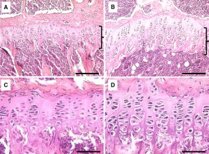
Here is the growth plates under LSJL(I provide a more detailed analysis here):

LSJL growth plates

“H&E-stained sections of the growth plate in the proximal tibia. a Growth plate of the contralateral control. The bracket denotes the growth plate. b Growth plate (bracket)
of the loaded tibia. c Proliferative and hypertrophic zones of the contralateral control. d Proliferative and hypertrophic zones of the loaded tibia. Bars a, b 100 micro-m; c, d 200 micro-m”

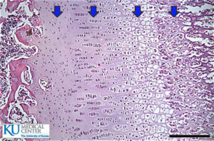
Here’s a baseline growth plate with similar colors:

resting-zone

It’s difficult to say exactly what is going on in the growth plates of the control and LSJL-loaded version but what is clear is that the micro-environment of the two bones is dramatically different.  The LSJL loaded growth plate has much more bone marrow and many more osteoclasts(the white spots; although those spots could also be adipose tissue).  The increase in bone marrow and loss of bone trabeculae would be more enabling for micro-growth plates.  Thus, LSJL could create a more favorable microenvironment for micro-growth plates.

“Histological analysis revealed that knee loading increased the height and the cross-sectional area of the growth plate in the proximal tibia. First, the total growth plate height was increased by 19.5% (175 ± 25.6 micro-m in contralateral control; 210 ± 18.1 micro-m in loading) including the heights of the proliferative zone and the hypertrophic zone. In particular, the height of the hypertrophic zone was extended by 33.6% (48 ± 4.6 micro-m in contralateral control; 65 ± 3.4 micro-m in knee loading). Note that the height ratio of the hypertrophic zone to the growth plate (HZ/GP) was significantly increased, whereas the ratio for the proliferative zone (PZ/GP) was not altered”

“the cross-sectional area of the growth plate was increased by 30.7% (0.263 ± 0.108 mm2 in contralateral control; 0.344 ± 0.095 mm2 in knee loading)”

“At the cellular level, the numbers of chondrocytes were increased in the total growth plate and the hypertrophic zone by 28.5% and 46.3%, respectively. In the proliferative zone, however, no statistically significant difference in the numbers of cells was detected”

“the height of individual chondrocytes in the hypertrophic zone was elevated in the loaded side (16.3 ± 1.67 micro-m) compared to the control side (13.0 ± 1.45 micro-m)”

“oscillatory loads laterally applied to the knee not only induce anabolic responses but also lengthen the femur and the tibia.”<-Interesting that they do not state the necessity of an existing growth plate in this statement although admittedly this is not strong evidence.

The total length increase in the growth plate was more than the sum of the increases in the proliferative and hypertrophic zones, indicating that other regions such as the resting and calcifying zones were also affected“<-This is huge as the resting zone is where chondrocyte progenitor cells are derived.  If LSJL can induce mesenchymal stem cells to become chondrocyte progenitor cells than it can create new growth plates.

“Because the cross-sectional area of the growth plate is significantly increased with knee loading[the growth plate is wider], the data support that the bone-lengthening effects are not limited only to the lateral or medial loading site. At the cellular level, the number of chondrocytes in the hypertrophic zone was increased together with their cellular height. Our results are consistent with the notion that dynamic tensile and compressive loads stimulate and suppress longitudinal growth, respectively”

“In knee loading, the rate of lengthening with 0.5 N loads (peak-to-peak) was 0.1% per bout (femur) and 0.1% per bout (tibia) for 5-min loading per day.”

“both loaded and contralateral hindlimbs increased in length in the tibia.”

LSJL Studies 1: Osteogenic potentials with joint loading modality

I will be going over all the LSJL studies to see if I missed anything or to find new insights.

Osteogenic potentials with joint-loading modality.

Here’s the paper: osteogenic LSJL study.
“Osteogenic potentials with a novel joint-loading modality were examined, using mouse ulnae as a model system. Load-induced deformation of rigid bone [generates] interstitial fluid flow and stimulate osteogenesis. However, in most of the previous studies, loads were applied to cortical bone. In the current study, we addressed the question of whether deformation of the epiphysis underneath the joint would enhance bone formation in the epiphysis{New bone formation in the epiphysis can increase height if the bone is added at the longitudinal ends} and the diaphysis. We applied lateral loads to a mouse elbow. Compared to the no-loading control, 0.5-N loads, applied to the elbow at 2 Hz for 3 min/day for 3 consecutive days, increased the mineralizing surface (two- to threefold), the rate of mineral apposition (three- to fivefold), and the rate of bone formation (six- to eightfold) in the ulna. Strain measurements indicated that strains of around 30 microstrain{30 microstrain is extremely low according to mechanostat theory}, induced with the joint-loading modality, were under the minimum effective strain of around 1000 microstrain, which is considered necessary to achieve strain-driven bone formation. To evaluate the induction of fluid flow with the joint-loading modality, streaming potentials were measured in separate experiments, using mouse femurs ex vivo. The streaming potentials correlated to the magnitude of the load applied to the epiphysis, as well as the flow speed in the medullary cavity.  Joint-loading [induces] osteogenesis, through a mechanism that involves the induction of fluid flow in cortical bone.”

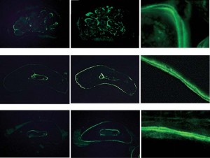
loading versus loaded LSJLColumn 1 is unloaded.  Column 2 and 3 is LSJL loaded.   It’s hard to tell in this pictures if LSJL created any gaps in the bone where new growth plates could form.  The diagrams are not in enough to tell to notice the formation of any microgrowth plates at least in figure 2a.  Calcein staining was used in these studies which detects mostly Ca2+ and Mg+ so it cannot distinguish between potential micro-growth plates and regular bone.

Here’s what the text had to say about the above diagram:

“Cross-sections of the ulnar shafts of control (no loading) and joint-loaded mice. The zoom images on the far right show double calcein staining, where the confined area constituted bone newly formed in 4 days. A Section of the metaphysis (trabecular bone) 1 mm from the loading center. The light staining outside the periosteal surface is collagen autofluorescence in a tendon of the triceps. B Section of the diaphysis (cortical bone) 2.5 mm from the loading center. C Section of the diaphysis (cortical bone) 4.5mm from the loading center”

“Trabecular bone in the epiphysis is less stiff in the lateral direction than in the axial direction and, therefore, lateral loads to the elbow may effectively deform the epiphysis of the ulna.
Deformation of the epiphysis may then induce fluid flow in the ulnar diaphysis in cortical bone, and load-induced fluid flow may enhance bone formation in the epiphysis{and possible stimulations of the epiphysis could be such as to spur new longitudinal bone growth} and the diaphysis.”

14 week old mice were used.  “3min per day for 3 consecutive days. The loading force was sinusoidal, at 2Hz, with a peak-to-peak amplitude of 0.5N.”

“The measured intramedullary streaming potential (f1, in mV) correlated to the magnitude of the applied force, according to the equation: f1 = 7.3 ¥ F (r2 = 0.92)”

“the magnitude of the streaming potential in the medullary cavity is proportional to the lateral load applied to the joint and the speed of fluid flow.”

If you look at figure 1a you can see that the device used is a lot like a C-class clamp with the nylon screw.

“The tip of the loader had a contact area of 4 mm in diameter. In order to avoid local stress concentrations between a joint and the loader, the surface of the loader was covered with a silicon rubber sheet.”

It should be noted by analyzing Table 1 that LSJL increases the bone formation rate of bone near the periosteum than trabecular bone.   Since the periosteum is partially involved in growth plate formation this is not necessarily a bad thing and since trabecular bone is still stimulated it still means that LSJL stimulates all areas of the bone and that LSJL could target any area of the bone that could be required to be targeted for neo-growth plate formation.

“[With axial loading], the force required to elevate the rate of bone formation is reported to be 2.3 N . With the joint loading modality described here, bone formation was enhanced by loads as small as 0.5 N.”<-So LSJL requires about 21% as much load to stimulate the bone as axial loading.  Let’s say hypothetically, that 1000lbs of axial loading could stimulate neo-growth plate formation.  LSJL would only require 210lbs.  Although, the rate of bone formation is irrelevant to what we’re looking for as we’re looking for longitudinal bone formation via neo-growth plates and that may be a result of stimulus that is unique to LSJL that axial loading cannot provide.

“The cross-sectional images of the ulna, together with the data on bone strains, support the notion that enhanced formation of cortical bone was an adaptive response to mechanical stimuli rather than a response associated with wound healing. First, the histological sections clearly showed double-labeled staining on the periosteal surface, with no indication of woven bone, which would frequently be formed in the process of wound healing. Second, unlike the four-point bending modality, where woven bone is formed underneath soft connective tissues, due to bending moment or compressive stress”<-it may be better if LSJL did increase bone formation as a result of wound healing as that could indicate the formation of holes in the bone where neo-growth plates could form.  However, the lack of woven bone could mean that cartilage was formed instead of bone which would be very promising indeed.

“the speed of the intramedullary fluid flow induced by 0.5-N loads, applied to the knee, is estimated as 476micro-m/s.”

This study was published in 2005 whereas lengthening of mouse hindlimbs with joint loading was published in 2010 so they were not yet aware of the lengthening effects.  And the load was only applied for 3 days which is not a lot of time for bone lengthening to occur.

I did email the author to try get length data but I don’t know if he’ll respond.