Author Archives: Senior Researcher

Stem Cells Implanted Into Bone For 12 Inches Of Height At A Clinic In China With South Korean Machine

Background On Sam Snyder

I contacted Sam in the early months of the the website’s development to ask about his own opinion on Tyler’s blog and what he felt was the direction of potential height increase research. Of all the bloggers in the internet who has ever done any type of extensive, serious review on Tyler’s HeightQuest.com website, it has been Sam Snyder, who talked about the research we might see in the decades ahead on what might be created biomedically for use. I have mentioned Sam’s name in a few past previous posts.

Something I found today which is really interesting is that the blogger Sam Snyder, who I have actually contacted in the past wrote a post back in July of 2011 which suggests that someone he knew was offer a chance to get their height increased by 30 cms from getting stem cells injected into their bones using a machine from South Korea and at a clinic in China.


Growing Taller with Stem Cells in the Future

Posted on July 15, 2011

Several months ago I wrote a post titled Stem Cells for Height, which outlined the possibility of someday increasing height through advances in biotechnology. I was recently contacted by someone who was offered the opportunity to increase their height by having stem cells injected into their bones at a clinic in China using a machine from South Korea. The procedure promised a height increase of up to 30 cm in adults! It would be nice if this technique worked, but I remain skeptical. The last time I checked, the website for this clinic had gone offline, so maybe it’s too good to true.

Articles like this are why I’m fairly skeptical of stem cell treatments that aren’t FDA-approved:

• Health Experts Warn of ‘stem cell tourism’ dangers

Regenerative medicine researchers say that patients could lose their money and have no recourse to get a refund. Patients might even get cancer from unrestrained stem cells. There still may be the possibility that stem cell clinics in China will develop successful treatments while American stem cell research proceeds at a slower pace due to FDA regulations. I’m not really sure what the right answer is. The regenerative medicine professors are skeptical of places like Beike Biotech, but that company seems to be conducting legitimate research (though admittedly I haven’t read their published papers):

• Beike Biotech Featured Publications

Legitimate stem cell therapies are still a major component of the future of medicine. Two major companies in America that are conducting clinical trials of stem cells are Geron and Advanced Cell Technology. Geron is conducting a trial of stem cells to heal spinal damage and Advanced Cell Technology is conducting experiments using stem cells to treat eye problems like macular degeneration. Bone marrow stem cells are also used to treat cancer. I know that other researchers are working on creating stem cell treatments to heal the heart muscle. Researchers have also successfully used tissue engineering to create new bladders and tracheas for patients. Anthony Atala of Wake Forest University has grown a kidney using tissue printing, but printed kidneys are still years away from being given to human patients.

Mesenchymal stem cells are also being used for bone healing in experiments, though I haven’t heard anything about being able to use them for the purposes of increasing height. Here’s a story about accelerating the healing of injured bones:

• Bone Growth Accelerated with Nanotubes and Stem Cells

Thousands of clinical trials using stem cells are being conducted or have been conducted. This is a list of clinical trials in the USA that are testing stem cell treatments:

• Stem cell clinical trial search results on ClinicalTrials.gov

As for increasing height using stem cells, I wish it was true. There are legitimate stem cell treatments out there, but unfortunately I’m not familiar with any that can increase adult height.

Updated 4/21/2013

The post he referenced at the beginning of the post was written in March of 2011


Stem Cells for Height

Posted on March 29, 2011
I recently read a demoralizing answer to a question in the following thread on Quora:

The top answer comes from a man who is barely five feet tall and experienced many setbacks in life. A growing body of research indicates that taller men have better lives on average. They have:

  • better career prospects
  • more frequent opportunities for romance
  • increased intelligence
  • happier mood
  • longer lifespan

Harvard economics professor Grew Mankiw, who himself is over six feet tall, wrote a semi-serious paper arguing in favor of a height tax. If economists truly believe in taxation that redistributes wealth to people who have poorer circumstances in life, that means they should examine the possibility of tax increases on tall men – or at least tax breaks for shorter men. The height tax is a politically untenable topic and is probably unnecessary in the long run, but evidence tends to suggest tall people have better lives.

Most people who provided counsel to the Quora respondent focused on short-term solutions (moving to a place with a population that has a lower than average human height) or long-term solutions (waiting for a technological singularity and then uploading the brain into a better body). Controversial limb-lengthening surgery also exists for the purposes of increasing height. The downsides are its expensive cost, the long recovery process, and the unimpressive results.

Stem cells are a promising medium-term solution. Stem cells could restore the body’s bones and muscles to a youthful state and allow for their expansion in a growth phase. They could also turn back the clock on the pituitary gland and cause it to produce excess growth hormone. This would be a delicate process to deliver the benefits of height without causing the health complications associated with gigantism and acromegaly.

Developed countries have lots of regulations on stem cell treatments and for good reason. They want to prevent people from wasting their money on scams or getting cancer from untested therapies. The good news is that some developing countries (especially in Southeast Asia) have fewer moral, ethical, and regulatory barriers to researching stem cell treatments. A country like China that has an excess of males who want to increase their mating prospects is prime territory for the development of height-increasing stem cell therapies. – Updated 11/27/2011

Personal Opinion

It is interesting that Sam mentions the company Shenzhen Beike Biotechnology Company because a source I found which discusses the issue over the ethics and moral implications of using stem cells is brought up in a journal by Boston College entitled “Ethical Concerns in the Emergence of Stem Cell Therapies by Benjamin Schanker

“…There may eventually come a day when organs or limbs can be manufactured, neuronal growth stimulated, or bone growth and height increased using stem cells. These further advances will certainly raise questions around issues of what it means to be a human being…”

This guy also states the possibility that maybe one day stem cell technology will allow people to increase their height.

The thing about this post is that Sam Snyder is claiming that there is some device in a clinic in China which was made or manufactured somewhere in South Korea which can implant stem cells into the bones of a person leading them to become upwards of even 12 inches taller.

I would love to hear from Sam to see who this person he knows is and whether that is any real evidence that this device exists somewhere.

I am in South Korea myself and plan to take a trip to China sometime soon so maybe I can do more research and find this actual device.

Increase Height And Grow Taller Using Pleiotrophin

I found an article recently which stated that a completely new compound which I have never heard of has the ability to increase the amount of stem cells taken from human blood and bone marrow. Personally I believe that one of the best approaches and paths towards alternative height increasing ideas is to study more on potential compounds that can increase stem cells. This compound called Pleiotrophin is very new to me so I asked the question “Can we potentially inject pleiotrophin in our cartilage or bones to induce chondrogenesis and cartilage formation, and thus increase in height and make us taller?

The article is entitled “Newly-Identified Growth Factor Promotes Stem Cell Growth, Regeneration” which I found from DukeHealth.org which is some website affiliated with Duke Medicine.

This compound seems to be a recently discovered growth factor that has the function of “stimulating the growth and regeneration of hematopoietic  stem cells in vitro and in vivo.” The new growth factor has potential since one of the biggest challenges with traditional applications of stem cells is the fact that stem cells seem to not proliferate very easily and the quantity of stem cells available is very little. It seems that the stem cell researchers have agreed that the ultimate source for stem cells for people who need transplants (ie. people who have cancer and can not longer use the chemo & radiation approach) would be from umbilical cord blood. The amount of umbilical cords are obviously extremely limited and there has not been any type of growth factor that is good enough in expanding the number of stem cells from the umbilical cord. When the researchers injected lab rats with pleiotrophin they noticed that the amount of stem cells that were coming from or produced by the bone marrow increased 10 fold. So obviously this compound might have some very important applications. However there is some worry that this compound might also cause some cells in the body to turn malignant although so far so results show this to be the case.

So I looked through PubMed and dug up as many useful and relevant studies as I could find to see what is the effect of pleiotrophin on bone tissue and cartilage tissue, if any. These are the studies that I managed to find.

Study #1: Pleiotrophin inhibits chondrocyte proliferation and stimulates proteoglycan synthesis in mature bovine cartilage.

Pleiotrophin turns out to be a type of regulated protein that is found commonly in the cartilage of fetuses but not developed mature cartilage. It binds to heparin. When the proteoglycan from the medium of mature chondrocytes are removed and treated with this compound, the synthesis of proteoglycan increases by 3 X. When the same pleiotrophin is added to the chondrocytes of fetuses, there is no increase in the level of proteoglycan synthesis. It seems that pleiotrophin seems to be able to control by inhibiting the growth of chondrocytes. When FGF and heparin was used, they could not reverse the effects of pleiotrophin. Upregulation of the mRNA for the proteins biglycan and collagen type II are also seen in the treated mature chondrocytes. It was noticed also that glycoaminoglycan concentration was increase multiple fold.

The researchers concluded that “These results establish that PTN inhibits cell proliferation, while stimulating the synthesis of proteoglycans in mature chondrocytes in vitro, suggesting that PTN may act directly or indirectly to regulate growth and proteoglycan synthesis in the developing matrix of fetal cartilage.
Study #2: Effects of pleiotrophin, a heparin-binding growth factor, on human primary and immortalized chondrocytes.

It seems that this compound is expressed in mature cartilage in the early stages of osteoarthritis. However it seems that if you stimulate chondrocytes with the pleiotrophin for a long enough time you reduce the mRNA of VEGF as well as Nitric Oxide production. The other big part is that the mRNA for MMP-1 & MMP-13 were reduced, while the inhibitors of the MMPs, something called TIMP-1 & TIMP-2 were induced, which suggest that it could be very useful for chondrocyte formation. It was concluded that PTN is a autocrine growth factor in cartilage which has a role during the formation of osteoarthritis when the chondrocytes start to aggregate together.

Study #3: Pleiotrophin, an embryonic differentiation and growth factor, is expressed in osteoarthritis.

This compound is said to be “expressed in mesodermal and neuroectodermal cells during development, but rarely in adult tissues“. The researchers wanted to see whether PTN would show up in the synovial fluid of joints which have osteoarthritis. It seems that this growth factor has roles in cancer development, in chondrocyte differentiation, and during embryonic growth. The most interesting conclusion is that during early stages of OA, the mRNA for this peptide is expressed. While some people might think it is a bas compound, the researchers conclude that “PTN might be involved in cartilage repair in OA, in particular, in earlier stages

Study #4: Expression of pleiotrophin, an embryonic growth and differentiation factor, in rheumatoid arthritis.

It seems that PTN was strongly up-regulated in synovial tissues from patients with rheumatoid arthritis while the up-regulation for people with osteoarthritis was much less. It also stimulated the proliferation of cultured human synoviocytes and in dermal fibroblasts VEGF. “Proinflammatory cytokines enhance the expression of PTN. Thus, we propose that PTN is a further paracrine angiogenesis and growth factor for synovial cells

So what did I find and conclude about this compound? Does it have any type of pro-chondrogenic potential or anabolic properties?

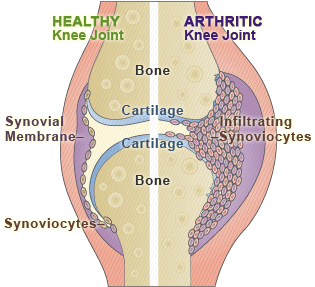
SynoviocytesAs a growth factor, it is definitely anabolic and seems to have some qualities that make it very useful.

It seems to cause inflammation but inflammation is just the body’s immune system trying to attack a foreign object.

This compound is seen in both of the main types of arthritis. The general guess is that it is not expressed to start to damage the cartilage, but it is actually used to heal the cartilage during the early stages of arthritis at least.

It is said that this compound inhibits VEGF, NO, MMP-1 and MMP-13, all of which we have concluded at some point is bad for overall cartilage health.

This compound was shown to increase GAG and Proteoglycan concentrations in mature cartilage chondrocytes and medium/matrix in vivo and in vitro.

I would guess that injection of this compound would NOT cause arthritis since it seems to be to repair cartilage. If you injected it, the matrix of cartilage might generate more GAG and make the cartilage more robust and full.

There is a slight change that this compound can be used to keep cartilage healthier and slow down growth plate cartilage ossification.

Cosmetic Surgery Mistake Reveals That Stem Cells With Hydroxyapatite Causes Differentiation Into Bone Tissue

While I was looking for sources yesterday which showed how stem cell research can one day be applied towards height increase I found a rather interesting story from the website for the The Daily Mail UK entitled “Botched stem cell facelift leaves woman with bones growing in her EYES 

The story is very much like what would be found in the tabloid stories in the US based Sun or Star Magazines but it did contain a small seed of useful information which we as height increase researchers might be interested in.

It seems that this women who went to a cosmetic surgeon in Beverly Hills noticed that her eyelids seemed to have been making a sort of “clicking” sound. She felt extreme pain when opening her eyelids. After going to another cosmetic surgeon to ask them what is the problem, it was found that she had bone tissue growing from her eye socket area.

From the article in the link…

“Dr Wu, of The Morrow Institute in California, told the magazine that when he first heard the woman’s complaint, he wondered if she was imagining things. 

But after painstaking six-hour surgery, he dug out small chunks of bone from her eyelid and the surrounding tissue.”

The clicking was two bone fragments that were scraping against each other. I would guess that somehow the injected stem cells that was taken from her stomach area turned into pieces of bones.

The researchers believe what happened was that the stem cells taken from the patient’s body reacted with a type of previous cosmetic surgery implantation, something known as dermal filler. The dermal filler has calcium in them. The dermal filler was added at the same time that her stem cells were added. The dermal filler was supposed to help remove wrinkles. The actual calcium is the calcium crystal hydroxyapatite

“During the procedure, cosmetic surgeons used liposuction to remove some fat from the woman’s abdominal area. They then isolated the stem cells.”

It seems that they took some adult adipose derived stem cells from the fat/adipose tissue around their stomach region. The tissue was filtered and purified to leave just the stem cells behind. The stem cells were injected into areas of the face to make the skin area look younger and healthier to produce what should have been new skin tissue and increase cell proliferation.

The 2nd cosmetic surgeon believed that the hydroxyapatite from the dermal filler got in contact with the stem cells which were injected into the face at the same time. Somehow the calcium crystal caused the adipose derived adult stem cells to start to differentiate into the osteogenic lineage.

The thing that is useful to know is the formula below

  • Calcium Crystals Hydroxyapatite would cause adult adipose derived stem cells to differentiate into the osteogenic lineage formed bone tissue. 
  • Hydroxyapatite + ADSCs = Bone

A Study Of Mesenchymal Chondrosarcoma – A Reply To Another Person Who Argues That Epiphyseal Hyaline Cartilage Never Disappear

I recently found from typing in the phrase “stem cell height increase” into Google a forum message entitled “The Science of Increasing Height” from the forum ScienceForums.Net

The post who goes by the name HeightAudodidactic started the post back in August 2010 with this segment which I will post below…

“I’ve done a lot of research on height increase and it’s hard to get information that’s not from laymen or people who are so advanced they’ll just blow you off(cartilage and stem cell researchers).

I can prove that it’s possible to increase height on every level. Long bones can experience appositional growth (there’s been studies done on runners showing increase in periosteal width unless the increase is due to thicker periosteum people being more successful runners) and short, flat, and irregular bones have periosteum covering longitudinal directions such as calcaneus, pelvis, bone of the skull, spine, etc. Therefore it’s possible to grow taller that way.

The epiphyseal cartilage does not completely mineralize. I have not seen microscopic slides of this but not because I have not been looking. However, a texture of whiteness can be seen in the epiphysis that’s around the same texture of the articular cartilage. I have read textbooks that state that a layer of hyaline cartilage remains in the epiphysis as well. Endochondral ossification can occur from a lot of places. See the study called: Reappraisal of mesenchymal chondrosarcoma… If a hyaline cartilage line exists in the epiphysis then growth could be enhanced in that manner either through extraneous stem cell injection or by trabecular microfracture in the epiphysis (which contains red bone marrow which contain stem cells). Also, the fibrous capsule contains cells that have the ability to differentiate into chondrocytes and studies on lateral loading increasing height (see study: lengthening of mouse hindlimbs with joint loading). Lateral loading does cause shear strain on the fibrous capsule And the fibrous capsule leads almost directly into the hyaline cartilage growth plate line.

I was wondering if I could get maven’s opinions on height growth and the topics I mentioned above.”

I wanted to talk about this poster’s points because some of what he/she was saying does make a lot of sense to any height increase researcher who is at a higher level of understanding. They claim that they can prove that height increase can be achieved at every level. They also wanted someone else who understood enough about the science and biology of the human body to sort of argue with them on whether they were right or wrong.

The task I felt was something that I should try to either rebuttal, analyze, discredit, or agree with. So first I wanted to focus on the highlighted parts because those are the point which I felt is the most unique and original, ideas put forth which I have not seen before.

Analysis #1:

The first thing that this person says is very controversial already. They say that they can prove that growth is possible. The claim that runners have wider long bones from looking at the cross section of the tibia/long bones in general might be true since appositional growth is something that does occur. Due to Wolff’s Law, the bones will from constant loading start to remodel the bone mineral density and the bone width as a response to the external stimuli. His comment that the periosteal part (outer part) getting thicker is right, and it just might be that the thickness is due to the periosteal appositional growth (where periosteum get thicker). However we must remember that the way that appositional growth even works is to grow bones on the inner germinal layer that pushes in the inward direction. The bones do get thicker on the outside, but the layer on the inside actually breaks down as calcium minerals get adsorbed into the blood in the cavity or get resorbed into the trabecular bone region. The direction of bone growth is from outside inward. This means that that net thickness of the bone doe snot change.

His next few claims are the ones that I would definitely contend with, and I am sure almost every person who has ever gone through medical school would contend this person’s claim as well. There is no hyaline cartilage layer left. I have actual X-Rays of my own knees from 4 years ago and from careful inspection, I do not see any line. Even the epiphyseal line that is supposed to result when the last bit of cartilage ossify seem to disappear as well over time. This guy talks about how he has not seen slides and that is the mistakes. I have seen the slides of at least two people in my life who were adults, one of them being mine. In both cases the X-Rays show that the long bones like the femur have absolutely NO line or white tint at the region where the growth plates used to be. There is just nothing there.

If this person’s argument in the beginning is challenged and disproved, which I think I have already, then all the arguments that they made after the first claim holds no water in logic. He talks about stem cell injections and trabecular microfracture in the epiphysis, which do make some sense and possibly work if there was a thin layer of cartilage still available, however there is no layer of hyaline cartilage left. It is all gone.

This guy uses the term ‘fibrous capsule’ and I would assume that the term is referring to the inner part of the overall long bone, like a femur. The bone is an enclosed area that encapsulates the marrow and progenitor mesenchyme inside. he talks about the inside of the fibrous capsule having cells that differentiate into chondrocytes and that it true. His mention of LSJL and the fact that it would cause the shear stress that leads to more stem cells that go towards a layer of hyaline cartilage cells and then start differentiating into the chondrocyte lineage, (although I state that he was wrong about this because the assumption is always that there is some chondrocyte-like layer still around where the growth plates used to be, but I challenged and disproved the initial assumption)

So it seems that this person got what they wanted, which is a somewhat educated response to their question.

For me I guess the most useful thing that this guy/girl showed was a study that they specifically named “See the study called: Reappraisal of mesenchymal chondrosarcoma…”. I found it, and wanted to look at the science of this type of cancer to see if it might have something useful we can learn from and take away for future research.

Reappraisal of mesenchymal chondrosarcoma: novel morphologic observations of the hyaline cartilage and endochondral ossification and beta-catenin, Sox9, and osteocalcin immunostaining of 22 cases.

This type of rare cancer is said to be a round cell and hyaline cartilage tumor. Researchers wanted to see what would happen if they put SOX-9, Beta-Catenin, and osteocalcin in people who had this type of cancer. The tumors were examined and some things were found.

It is noticed that in the tumor, there are cartilage cells that are going through a similar process as if they were going through endochondral ossification. Cells positive for osteocalcin were found. There seem to be at least two types of sarcomas defined in this abstract, the mesenchymal chondrosarcoma, and the small cell osteosarcoma which seem to be formed from small round cells and turn to osteo-like tissue.

The sentence that the guy was referring to on why they say that cartilage can be regrown or regenerated is probably the one below…

“Mesenchymal chondrosarcoma demonstrates centrally located hyaline cartilage with a linear progression of chondrocytes from resting to proliferative to hypertrophic, which undergoes endochondral ossification, recapitulating growth plate cartilage and suggesting that this component of mesenchymal chondrosarcoma may be a differentiated (benign or metaplastic) component of a malignant metastasizing tumor…”

This seems to me to mean that if a person developed this type of cancer, there will be hyaline cartilage that is formed in the cavity in the middle of long bone which has all of the same types of chondrocytes as natural growth plates. It says that growth plate cartilage is recapitulated, which implies that the cartilage can be regrown over again from the advent of this cancer.

Implications For Height Increase

I am not sure at this time how to interpret this abstract and whether I am even reading the abstract correctly and understand it like I am supposed to. It seems to show that for people who have this rare condition known as msenchymal chondrosarcoma who are adults, tumors in the center of long bones develop. This tumor are NOT the same as the osteosarcoma developed by small round cells which ultimately form bone tissue. This tumor seems to consist of a linear progression of chondrocytes that look very much like the histology of natural growth plates.

This might mean to show that for people who have this disorder, they have newly formed growth plates but I am very cautious on this claim because I still understand very little of what is really going on with the bone tissue. Even if growth plate structure is noticed in the center of the bone, it does not mean that the growth plate has the strength to expand to a degree to pull the cortical bone apart to lead to renewed longitudinal growth.

So more research needs to be done.

Polyacrylamide and Alginate Hydrogel Used As Scaffold Is Alternative To Autologous Chondrocyte Implantation

This study I found suggest that instead of the traditional approach in tissue engineering where the scaffold where the stem cells or growth factor is placed is made of not very strong or elastic in terms of material and machanical properties, it might be possible to add a scaffold made out of polyacrylamide and a type of alginate hydrogel which has much better mechanical and material properties.

Update – Link seems to NOT work: New hydrogel may help cartilage regeneration research

Link #2 – New ‘hydrogel’ shows promise for cartilage repair in proof-of-concept trial

This link is nothing new to me but shows that in terms of the development of certain types of biomedical research into actual application, it has taken a long time. For the last decade or so scientists and researchers around the world have been using a stem cell w/ scaffold combination to regrow bones and cartilage. Only now has this news been reported, and from John’s Hopkins University. Like the article states, many defects seen in the articular cartilage in humans have been traditionally fixed using the microfracture surgery, where a small drill is done at the defect area. The marrow inside pours out, forming a layer of fibrocartilage which is just not as good as the hyaline cartilage that was there before. This scaffold idea which is supposed to be a ‘breakthrough” is really not. It might be that researchers at JHU have been trying out thousands of scaffold composition or formulation combinations before they finally found a good enough hydrogel/scaffold/alginate combination for stem cells to really grow. The combination of the idea of stem cell in scaffold along with microfracture surgery is not a big leap in understanding or application. I say that it was bound to happen eventually. The main thing I guess that is important is in showing that if you use the scaffold and microfracture in combination, it is much better than just going with the microfracture sugery route along.

The results are…

After six months, the researchers found that hydrogel recipients had new cartilage filling 86 per cent of their defects, on average, compared with just 64 per cent for patients with microfracture alone.

In addition, patients who received the hydrogel implant reported a greater improvement in their levels of knee pain.

Study #1 – Highly stretchable and tough hydrogels

Summary – Hydrogels has been used in many types of applications but they are not very good due to having certain types of mechanical properties. They are not very elastic and break after they are stretched even slightly. Compared to other materials like cartilage and rubber, the hydrogel does not have a very high fracture energy, making them much more brittle. At the current moment, multiple researchers are looking for ways to make a type of hydrogel that is much stronger and more elastic. There are already some synthetically made hydrogels that have a fracture energy of 100–1,000 J m^(-2) which is up to 2 magnitudes greater than average. 

However, for this study, the researchers show that they have developed a new type of hydrogel that is much more elastic and have much higher fracture energy and stretchability. They report the synthesis of hydrogels from polymers forming ionically and covalently crosslinked network

These new synthetic hydrogels can stretch up to 20 X their length, have fracture energy around 9000 J/m^2 and be composed of 90% water.

They state that the gels’ toughness to the synergy of two mechanisms:

  1. crack bridging by the network of covalent crosslinks – Furthermore, the network of covalent crosslinks preserves the memory of the initial state, so that much of the large deformation is removed on unloading.
  2. hysteresis by unzipping the network of ionic crosslinks – The unzipped ionic crosslinks cause internal damage, which heals by re-zipping.

Implications For Height Increase Application

I personally think that the best and most likely alternative to the limb lengthening surgery is doing a type of synthetic tissue implant into the bone of a person. If we can create a strong enough type of growth plate in a scaffold formation, then it would definitely be a real alternative. This requires the understanding of stem cell mechanisms and tissue engineering practices. From tissue engineering practice, the use of scaffolds is very big. The use of hydrogels as scaffolds is very common, if not universal. I have been trying to figure out what types of hydrogels would be best to be used in combination with formed cartilage-bone tissue that is growing volumetrically.

As I stated in one of the most important posts on the website, we have been able to create growing cartilage tissue aka functional growth plates however I had stated that I worried that even if we implanted the cartilage into the body, they would not be able to withstand the loading by an adult human. This is why I felt that maybe we need to find a good enough transport system aka scaffold we can put not just stem cells in, but also expanding cartilage tissue. If the hydrogel scaffold is strong enough, the synthetic growth plate with strong & elastic scaffold combination being implanted would probably work, at least with multiple testing done.

From the study “Elastic, Superporous Hydrogel Hybrids of Polyacrylamide and Sodium Alginate“…

These hydrogels are distinguished from other porous hydrogels in terms of their pore sizes and the methods used to generate the pores. If any portion of a superporous hydrogel is exposed to water or aqueous fluids, fluid is immediately absorbed through the open channels to fill the whole space. This capillary-driven absorption mechanism helps dried superporous hydrogels to swell very quickly into a very large size.

These products are very resilient and resistant to compression and elongation. In their water-swollen state, elastic superporous hydrogels can be repeatedly stretched to almost twice their original length without breaking. These novel products may find applications in the development of drug and protein delivery systems, fast-dissolving tablets, occlusion devices for aneurysm treatment, scaffolding, cell culture, tissue engineering, water-absorbent pads, hygiene products (baby diapers, feminine pads) and many others.

It might be possible that we might use the Superporous hydrogels (SPHs) since they are made from hydrophilic polymers with so many pores and they also have the interesting property that the hydrogels EXPAND when they absorb water. It could be that the properties of Superporous hydrogels will be very good with the synthetic growth plate cartilage implanted since both will be expanding.

Mesenchymal Stem Cells Injected Into The Intervertebral Discs Does Increase Height and Restore Lost Height From Aging

This is sort of old news in terms of how advanced our research have become. Me and the other height increase researchers have known for quite some time that many types of growth factors will lead to height increase from the height lost from just the normal agin process. The most obvious one is the BMP-7 (aka OP-1) which was shown to increased the intervertebral disk height in White New Zealand Rabbits in.

It seems that a few other source around the internet have also picked on the idea and wanted to report that apparently from just a simple needle injection of the mesenchymal stem cells into an aging adult’s intervertebral disk, their lost height can be restored.

The article below was taken from a website called Healio Orthopedics Today

New research underscores potential for stem cell use in disc regeneration

Culture conditions affect the ability of mesenchymal stem cells to differentiate into intervertebral disc cells.

  • Orthopaedics Today Europe [Archives], Issue 5

VANCOUVER, Canada — Techniques that may employ mesenchymal stem cells to regenerate cells of degenerated human intervertebral discs showed that microenvironment plays an important role in cell proliferation, as does the cells’ ability to produce extracellular matrix.

Results of three laboratory studies and one using a rabbit model that investigated these areas were presented here during the International Society for the Study of the Lumbar Spine 30th Annual Meeting.

The concept of rescuing degenerative discs by injecting them with cultured mesenchymal stem cells (MSC) is not new, but more information is needed about the best MSC sources, culture conditions that support cell differentiation, and techniques that yield the greatest number of cells.

Me: I think we have reached the same agreement with the orthopedic surgeons. We can get height back from cultured MSCs however at this point, we just don’t know which mixture of growth factors and which source of MSCs would be the best and allow for the highest level of cell proliferation.

Also stated are these facts…

“Mesenchymal stem cells have the potential as an ultimate alternative in cell transplantation therapy for degenerative disc disease,”

“…found that within the disc environment, transplanted MSCs differentiated into disc-like cells, survived and proliferated, preserving disc structure”

“Disc height 26 weeks after degeneration was 90 ±8% in the transplanted group compared to 67 ±8% in controls…”

“MSC transplantation restored synthesis of a proteoglycan-rich matrix.”

“Our study has implicated the potential of MSCs to differentiate into intervertebral disc cells, which provides new information in MSC research”

Interpretation Of The Facts

The idea of doing transplants of cells as a type of therapy to treat degenerative dics disease is extremely possible and viable. The cells that are transplanted do differentiate into disk like cells, don’t die out, and do multiply thus maintaining the extracellular matrix environment of the annulus fibrosus and nucleus pulposus that they were injected into. This shows that the cells that are basically foreign objects seem to go along with how their environment is like and does not try to disrupt the collagenous and cartilagenous tissue content. The two groups in the experiment showed that compared to the controlled group, the rabbits that did get the injection got a significant amount of disk height back. The MSCs restored the synthesis of the matrix which has a lot of proteoglycan.

Furthermore, another group of researchers from John’s Hopkins University School of Medicine, the department of orthopaedic surgery at Thomas Jefferson University in Philadelphia, and the Injury, Repair and Rehabilitation Research Group at the University of Manchester, England states about their research…

“…investigated whether bone marrow-derived MSCs could stimulate the anulus fibrosus (AF) and nucleus pulposus (NP) cells they might eventually interact with during clinical application…”

“…Human NP and AF cells isolated from adult degenerative discs cultured in a commercially available MSC medium yielded higher glycosaminoglycan (GAG) content, which increased over time, compared to controls of NP and AF cells…”

“Co-cultured disc cells and MSC could aggregate and produce an extracellular matrix, and there is upregulation of the GAG content between the cells…”

“…degenerative disc cells may be stimulated in vivo by implantation of autologous MSCs to restore some of the disc properties…”

“given the proper microconditions, MSCs can express a phenotype like that of NP cells”

“Hypoxic conditions in combination with the alginate culture and the growth factor that we chose, which was TGF-ß, were really optimal for achieving an NP-like phenotype…MSCs cultured in alginate and pellet culture had the most chondrogenic characteristics, forming an extracellular matrix similar to that seen in vivo”

“…MSCs grown in monolayer in basic media transfected with SOX-9 differentiated and expressed type II collagen, aggrecan and SOX-9, but not type I collagen…”

“…Differentiation improved and there was high-level matrix production using TGF-ß1 in combination with SOX-9,…”

What we are seeing is just how powerful and potentially good the MSCs can be towards height increase at least for the torso region. The autologous implantations of MSCS can stimulate increased glycoaminoglycan content. They can also turn into the nucleus pulposus phenotype with the right environment. This can be achieved using a hypoxic induced environment with the growth factor TGF-Beta. The results is the formation of extracellular matrix that is similar to the annulus fibrosis and nucleus pulposus. The other idea is to put the SOX-9 gene in a vector to alter the MSCs to differentiate and produce the right type of compounds that would form the matrix. The type of compounds the MSCs would make are the Collagen Type II, Aggrecan, and possibly Hyaluronic Acid like glycoaminoglycans that we have seen in the matrix of ordinary hyaline cartilages.