Author Archives: Senior Researcher

Using Extracorporeal Shock Waves, ESWs To Stimulate Periosteum Osteoprogenitor Cambium Cell Proliferation In Non-Invasive Bone Tissue Engineering Application ( Big Breakthrough!)

Something which I have always guessed could really work in making people grow taller was the use of Extracorporeal Shockwaves. I had written before at least multiple posts about the possibility, showing the dozens of PubMed articles all suggesting that ESW therapy works in bone healing and bone growth, but also the possibility to cause chondrogenesis and chondrocyte proliferation. The post where I wrote about this idea was “New Proposed Height Increase Method Using BMPs And Extracorporeal Shock Wave Treatment, ESWT

For the Full version of the entire article, click HERE

Title Of Thesis: Non-invasive shock wave stimulated periosteum for bone tissue engineering

Kearney, Cathal (Cathal John) – Harvard University–MIT Division of Health Sciences and Technology.

Thesis (Ph. D.)–Harvard-MIT Division of Health Sciences and Technology, 2011

The cambium cells of the periosteum, which are known osteoprogenitor cells, have limited suitability for clinical applications of bone tissue engineering due to their low cell number (2-5 cells thick). Extracorporeal shock waves (ESWs) have been reported to cause thickening of the cambium layer and subsequent periosteal osteogenesis. This work proposes that ESW-therapy can be used as a non-invasive, inexpensive, and rapid method for stimulating cambium cell proliferation, and investigates the use of these cells for orthotopic bone growth. The response of periosteal cells to ESWs was evaluated using two different energy densities applied to either the intact femur or tibia of the rat. Just four days after application of ESWs, there was a significant 3- to 6-fold increase in cambium cell number and thickness. The most effective treatment of those tested was high dose ESW applied to the tibia. Immunohistochemical staining of the proliferated cells demonstrated osteoblasts and bone formation (osteocalcin stain); it also demonstrated extensive vonWillebrand factor expression, which reveals the vascular contribution to the proliferating cambium layer. In a rabbit model, ESW-thickened cambium layer cells were overlaid in situ on a porous calcium phosphate scaffold. At two weeks post-surgery, there was a significant increase in all outcome variables for the ESW-treated group when compared with controls: a 4-fold increase in osteoprogenitor tissue in the scaffold upper half, a 10- fold increase in osteoprogenitor tissue above the scaffold, and a 2-fold increase in callus size. The results successfully demonstrated the efficacy of ESW-stimulated periosteum for bone tissue engineering.

Analysis & Interpretation:

The thing about Ph. D programs are that they are very long and mentally intensive. What is well known is that for a person to be awarded a degree in graduate school, for say a Masters or Doctorate, they have to do more than just regurgitate the information that is already in the scholarly books, but create something new or answer some question which has not been answered yet. Also, Ph. D. Thesis tend to be more like novels since they are almost always over 100+ pages in length. This means that I will NEVER take that much time in analyzing every single sentence.

However I will explain why this MIT Thesis from 2011 is so important. It shows that we have finally discovered a real way to increase out height noninvasively, through the application of a type of vibration or wave to stimulate cell proliferation in an area of the body which can be stimulated since it still has progenitor mesenchyme-like cells which can differentiate into the type of cells we are looking for. 

From my analysis of just the Abstract this is the real deal.

Not only does this show finally the first real to way to increase our height, it also validates one of the primary ideas I have been having, which is that we might be able to manipulate the cambium cells in the lower periosteum layer to grow in thickness from proliferation. 

Increased thickness means the end of our long bones in our legs would be thicker, which translate to extra height.

If you are a true researcher, then I would suggest you go through the pages 40-52 where the Ph. D candidate goes into the introduction on the periosteum and the responses to extracorporeal shockwaves. The Thesis has results sections, but I would rather just focus on reading the “discussion” sections to see how the researcher is interpreting the data. I would assume that a person who has managed to make it through the Ph. D program in the hard sciences or engineering from MIT is intelligent enough to read and interpret the data they have received correctly, since they probably have been working on the problem for at least a couple of years.

They state …

“The focus of this thesis is the generation of bone orthotopically using ESW-stimulated periosteum in combination with a subperiosteally implanted porous cal- cium phosphate scaffold. ” (pg. 43 of 225)

This may mean that the person is only doing the research to make bones stronger and have greater bone mass density (BMD) to treat aged people with osteoporosis but it also shows that the results mean that applied the right way a subject can in theory gain a little bit of height increase.

Let’s look at this picture I clipped from page 44 which shows the diagram on how autologous chondrocyte transplant or autologous chondrocyte implantation (ATI) treatment would work.

autologouschondrocytetransplant


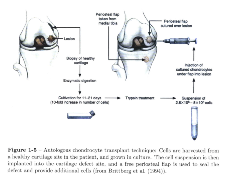
This is how the general implantation is supposed to work. However the technique here is specified for using a periosteal flap for a place to get chondrocytes. in using the periosteum. The rather sucky thing is that this guy did not decide to look at how feasible it would be to use periosteal tissue in cartilage tissue engineering and cartilage regeneration. They focused on bone growth.

From page 45 in the section 1.3.2 entitled “Thickening of Periosteal Cambium Layer”

Thickening of periosteal cambium layer

Previous investigators have reported techniques aimed at thickening the cambium layer of periosteal cells for use in tissue engineering strategies. O’Driscoll’s group injected TGF-#1 subperiosteally to stimulate periosteal proliferation and to initi- ate chondrogenic differentiation of the cambium cells (Reinholz et al., 2009). Simon et al. (2003) investigated the use of surgical techniques to stimulate periosteal cell proliferation in a caprine model. The authors performed surgical release of the pe- riosteum which resulted in cambium cell proliferation and subsequent bone formation within the proliferated layer 

From Section 1.4 on Extracorporeal Shockwaves…

Our supposition is to employ extracorporeal shock waves (ESWs) as a non-invasive and rapid way of safely and reliably stimulating the proliferation of the periosteal osteoprogenitor cells for use in orthotopic bone generation strategies after only a few days 

Importantly for this study, ESWs have been reported to stimulate periosteal osteogenesis following thickening of the periosteal cambium layer (Takahashi et al., 2004; Tischer et al., 2008). For medical applications, the peak ESW pressures lie in the range of 5-12OMPa; the rise time is on the order of 10-100ns; and the pulse width is on the order of 1ps (Chung and Wiley, 2002; Gerdesmeyer and Lowell, 2007).


We have to clear evidence from at least two studies that the ESW would work in making the bone ends thicker then.

Under Section 1.5 for Thesis Goals….

This thesis deals with a new therapeutic approach for regenerating bone orthotopi- cally. The emphasis is on growing bone on the cortical bone surface, which would have applications in bone regeneration in oral and maxillofacial surgery (e.g., alveolar ridge resorption), as well as in orthopaedics (e.g., osteolysis). The central hypoth- esis is that extracorporeal shock wave (ESW)-stimulated periosteum enhances bone formation in porous biomaterials implanted under the periosteum. The overall goal of this work is to demonstrate the efficacy of an ESW-thickened periosteum as an overlay on a porous calcium phosphate scaffold for the orthotopic growth of bone.

If we just skip to pages 72-75, where the Ph. D Candidate writes in the Discussion and Summary section…

Section 2.4

The objectives of this study were to determine the effects of energy flux density (0.15 vs. 0.40 mJ/mm2) and anatomic location (femur vs. tibia) on the periosteal cambium layer cell number. For all groups evaluated, cambium layer cell counts, and cambium layer thickness, were significantly increased following ESW treatment when compared to controls. The higher dose of ESWs resulted in a thicker periosteum compared to the lower dose (Group 3 vs. Group 4; t-test, p<.0001), which is consistent with previous findings (Tischer et al., 2008). However, the results presented here demonstrate a lower threshold for periosteal proliferation (0.15mJ/mm 2 ) when compared with a previous study that reported a minimum energy density of 0.5mJ/mm 2 was required for periosteal osteogenesis (Tischer et al., 2008). The data presented here show that a lower energy density, which reduces inflammation, can be used to stimulate the cambium cells. Tibial periosteum displayed a greater degree of cellular proliferation in response to ESWs compared to the femur (Group 3 vs. Group 2; t-test, p<.0001; tibia vs. femur). The larger amount of soft tissue surrounding the femur may cause more attenuation of the shock wave than occurs for the tibia. 

Conclusion:

From Sections 1 and 2, we have clear evidence that bone growth can rather easily occur from ESW application on the cambium layer of the periosteum. With higher dosages, at least to a energy flux density of 0.40 mJ/mm^2 the cell layer gets thicker. However the even better news is that the threshold for cell proliferation is lower than the results found from previous studies.

Section 3 is more interesting. The graduate student tried to figure out what types of cells are really found in the cambium layer and he says that the layer of tissue has mostly osteoblast-like progenitor cells, which can be labeled as mesenchyme and sort of “stem-like”. The problem for our goals is that the cells seem to favor and almost always take the path of differentiating into the bone cells, not cartilage cells. They note that previous studies seem to see the same thing, that the cambium progenitor mesenchyme go through the process of intramembraneous ossification, not endochondral ossification. When they checked to see if the cells left any Collagen Type II or Type X, suggesting chondrocyte production and chondrocyte hypertrophy, they found none.

For our guesses that the bone formation is from microfractures induced, the candidate says it is not likely, or that it could be periosteal elevation. What is most important is to show that vascularization is almost immediate after ESW treatment, which might explain why differentiation into osteoblasts are the main pathway goal than chondrocytes.

The reason for the vascularization and the intramembraneous ossification seems to be from two new proteins which I am not familiar with called SMAs and vWFs (VonWillebrand factor) which seem to be found a lot from endothelial cells.

This makes the candidate believe that the endothelial cells might be stem-like and multipotent differentiating only into bone cells when the ESW is applied causing vascularization.

At this point, we end Section 3 and the summary, and reach around page 95 of this 225 page Ph. D. I will probably try in a post later to go through this entire Ph. D and explain away the other sections to explain what all the technical jargon implies. The big thing to take away from this post are these two points.

  • ESW application even at a lower energy flex density does cause the periosteum to grow more bone from cambium cell thickness increase, which means we will grow taller by a little using this non-invasive method. 
  • The problem is that the ESW causes osteogeneis but not chondrogenesis, which is what we should really be focusing on to get grow taller by a much larger amount. Osteogenesis might increase our height by 1 cms maximum (a personal guess) but Chondrogenesis could lead to hypertrophy and multiple inches in height increase.

Medical Analysis On A 8 Feet Tall Iranian Giant, Siah Khan

I have talked about Siah Khan And Zech Devits before in the posts “The Development Of Gigantism Without Pituitary Abnormalities: Studying Zech Devits And Siah Khan, (Important!)” and “The Connection Between Matt McGrory, Siah Khan, Zech Devits, And The Tallest Filipino Leads To Proteus Syndrome And Much More (Important!)

This post is to focus more on what could be the genetic mutation which lead to Siah Khan. His case is one of the most unique, and most interesting of all giants I have studied. The abnormalities found on his face makes him so unique that there has really only been 1 case of his type in all of documented medical history.

The main resource I have to use was the study “Cranio-Spondylo-Tubular Dysostosis A Unique Historic Iranian Giant” –

Note: I do have a copy of the full PDF of the study somewhere on this website.

Author(s): Mohammad Hassan Sheikholeslami, Yousef Shafeghati , Zabihollah Ghorban

MD – Tele-fax: +98(21)44 633 283

Article abstract:

Severe overgrowth and tallness is very rare in human beings. The most common cause is gigantism due to the excessive secretion of the growth hormone, especially, before the closure of long bones’ epiphyseal growth plates. There are other rare disorders that are categorized on overgrowth syndromes. Herein, we report an extremely rare, or even perhaps a unique, patient from Iran. The clinical and skeletal findings were very unusual, with extensive clavarial, tubular, vertebral, ribs, and scapular overgrowth and synostosis. Indeed, the results of these abnormalities made a monstrous giant with a very tall stature. This is a unique case, which was living during 1912-1940 in Shiraz. The report’s information and photos, kindly supplied by Prof. Sheikholeslami. We evaluated thoroughly the findings together. Now we think; this is a very unusual case of its type, perhaps a Craniotubular Dysostosis Syndrome. We searched medical and clinical genetics literature, but did not find any similar case, having been reported before. So, to our knowledge, this is a unique case in the history of world medicine. We suggest to call this entity; “Siah-Khan syndrome” (after the patient’s name), or “ Ghorban-Sheikholeslami-Shafeghati Syndrome” (in honour of Prof. Ghorban who was recently has died, or “ Cranio-Spondylo-Tubular Syndrome”.

The other main website to see what Khan was like is from a website that is almost all in Arabic (click HERE)

Analysis & Interpretation:

Sometimes the doctors or researchers who study these giants are too busy taking pictures, and trying to handle all the press and media from having such a patient that they barely ever do any science or analysis on why this Iranian giant ever even existed. The other title given for the study was “A Unique Historic Iranian Giant “Siah-Khan Syndrome”, Report of an extremely rare or perhaps a unique case in the world from Iran”. The paper first states that this guy was born in the early 20th century to a normal family (father and mother and siblings are all normal). His birth shows that he was average sized. His growth spurt started around the age of 6 and by the time he was 9 years old, he looked like a guy who was 20 years old. Siah Khan started to develop deformities on his skull, face, arms, and legs and young kids started to become very scared of him. Eventually the family managed to get in contact with a Professor Ghorban in 1922. Khan was subsequently put in the hospital for care. He stayed there until he was 28 years old, when he died from pneumonia and sepsis in 1940. After his death, the professor felt his body was too precious and unique so didn’t bury it but put it up for display, where it is supposed to be presently in the main entrance of some university called Shiraz Medical Science University.

The paper itself is only 5 pages long and says that Khan was 259 cms tall and 250 kgs. If the numbers are true, that means Khan was standing around 8 feet 6 inches tall! It is also written that Khan is supposed to be mentally below average with a huge sexual apetite and could not turn left or right, or even stand up from sitting down. There is clear bony projections on the person’s face. 

They state….

Occipital bone growing backward.

– Frontal bone growth progressing to the forward and downward of the eyes as a thick wall limiting the field of vision just like a front porch.
– Large hands with long fingers.
– Large feet, very flat feet, bumping steppage.
– Great appetite as much as 3 to 4 people with good appetite.
– Looking like a strong, dry piece of thick wood and unable to move in different directions because of the fast growth of bones and early vertebral body fusion 

The height of the skeleton is 259 cm; the lengths of the upper extremities were 117 cm, and the lower extremities 125 cm. The very severe overgrowth in calvarial bones, clavicles, scapulae, long tubular bones, ribs, and pelvic bones are apparent. 

tibia porosityI personally took the liberty to clip and put the picture in the article of Khan’s only 2 X-rays, of his skull and his lower limb bones. It is noted that his tibia and also fibula was rather enlarged and sort of porous.

The physicians are right that there can be many different causes and reasons for gigantism, which is supposed to be labeled as any person who is 3 or more standard deviations above what the average height of people in their main group should be. After listing through what could have cause the gigantism and bony overgrowth on face, the Iranian researchers seem to suggest that Siah Khan suffered from something called Craniodiaphyseal dysplasia. As they say it…

Craniodiaphyseal dysplasia (CDD) is a rare craniotubular remodelling disorder with hyperostosis and sclerosis of the skull and facial bones, along with hyperostosis and defective remodelling of the shaft of the tubular bones. The epiphyses and metaphyses are only mildly affected.  

The other disorder which they sort of claim could be the cause is something called Craniometaphyseal dysplasia (CMD)

In Craniometaphyseal dysplasia (CMD), there is progressive facial dysmorphism with a broad osseous prominence of the nasal root extending across the zygoma. Autosomal dominant and autosomal recessive types of inheritance have been described. In general, recessive CMD is more severe than the dominant CMD 

The thing which the scientists at that time, and which we as height increase researchers can only say is that Siah Khan suffered from a very sever form of CDD or CMD, and his case is very unique. There was little blood work and no genetic studies done on him, since he lived in the the early 20th century. If he was born in the 21st century, we probably would have been able to help him out much more and be better and figuring out what exactly caused his condition.

siah khan 2

siah khan

siah khan

The Development Of Gigantism Without Pituitary Abnormalities: Studying Zech Devits And Siah Khan, (Important!)

In the last post I had found from forums off of other websites that there have been a few people who developed a type of gigantism without going through the normla pituitary abnormalities we are used to seeing. It seems that through certain genetic mutations, a few people have managed to develop gigantism.

The paper I would cite at the end of the previous post “The Connection Between Matt McGrory, Siah Khan, Zech Devits, And The Tallest Filipino Leads To Proteus Syndrome And Much More (Important!)” was entitled “A provisionally unique syndrome of macrosomia, bone overgrowth, macrocephaly, and tall stature

Abstract

We report a young man with intrauterine macrosomia, macrocephaly, and bony abnormalities. Excessive growth continued throughout infancy and childhood. Bone age was advanced. He developed contractures of the large joints and was confined to a wheelchair. Extensive laboratory studies, repeated on multiple occasions were all normal. Intellectually, he was normal. His near final height was 234 cm. The constellation of findings in this patient is at variance with previously described syndromes of tall stature. We postulate that excessive size and bone overgrowth in this young man is caused by a receptor/post-receptor abnormality involving a growth on/off mechanism at the cellular level.

I would take the 4 page full file from the forum thread HERE from TheTallestMan.com website. An expanded view of the 4 pages is available from the forum. For the analysis of the study go to the bottom of the post. There is actually also a whole treasure trove of studies and articles that a serious height increase researcher can mine from to learn more about how human growth occur.

11

121314

Analysis & Interpretation

The study that we find from 2005 was done on the giant Zech Devits who has passed away. He was measured (or maybe guessed) at a truly staggering 7′ 8″ tall. His large ears might have given off the fact that his height may be the result of excessive cartilage formation of hyperplasia.

From the abstract we learn that this patient had an above average sized skull, joints, joint curvature. HIs intellect seems to be intact. Besides the multiple issues the doctors and researchers might have found in his major organs, the skeletal structure showed mild contracture of the elbows and knees, the flaring of the long bones in the metaphysis region, and the curvature of the vertebrate aka thoracolumbar kyphosis. There was also thoracic dystrophy and diffuse bone dysplasia. What is really interesting to see from the charts of the blood tests is that the patient was not hypersensitive to the traditional growth factors which would cause overgrowth like insulin, growth hormones, or insulin like growth factors, IGF-1. At at the age of even an infant he was documented to have had spinal fusion. Around the age of 2 testing found that he was suffering also from severe spinal stenosis. At the age of 4.5 there was more signs of spinal atrophy.

Before even the age of 10, he was already almost 6′ 7″ (200 cms). Bone density decreased, myopia set in. From doing the bone maturity test using the Tanner method, his bone age around 9 showed that he was progressing at the average rate of most kids. His eyebrow and forehead developed, his ears grew out to be quite big, and his nasal bridge also increased. This suggest that the places of cartilage really decided to increase in size. Eventually his conditon got really bad and the people who were monitoring him decided to give him 200 mg/ week to speed up the bone maturation and fusion process.

From the discussion we would learn that while Zech was born prematurely, he was a big baby. The researchers note that his facial features resemble patients which have Sotos Syndrome or Weavers syndrome as well as Marfan’s Syndrome.

He had weakness in the upper body, moderate acne (which would suggest that his growth process of going through puberty since males start around 10-12 was normal for age range), and a very deep voice. This suggest again that his condition caused cartilage hyperplasia, since the pitch of one’s voice comes from how wide the cartilage of the windpipe (aka trachea)  and voice box (aka larynx) are. The wider the speaking windpipe and voice box, from say chondrocyte hypertrophy or proliferation, the deeper one’s voice.

What seems to be ever more interesting is from the photo in the article is that his head is very large compared to the average person. It is noted that he has supra-orbital ridges or what the common person would say is a very distinct, large eyebrow ridge which juts out. His hands and feet were large (size 22-23) but his fingers seemed to be relatively short. From X-rays done, it was found that he had most of the epiphyseal growth plates already starting to fused together even at the age of 12.5 from the estrogen-like hormone treatment, including the radius, femur, and tibia.

The conclusion the physicians who were trying to figure Zech’s condition out basically are not sure whether he was suffering from Weaver’s Syndrome or Soto’s Syndrome, but only that his asymmetrical nature of growth could be from Proteus Syndrome since Proteus Syndrome leads to asymmetrical overgrowth.

I would conclude this case of gigantism and over growth with a clip on the analysis the physicians gave on his condition and what might be the cause. At this point I don’t understand what they are talking about. What is clear to realize is that for Zech, he did not suffer from some type of hypogonadism or pituitary problem like most giants, but had some other type of disorder.

zetch

The Connection Between Matt McGrory, Siah Khan, Zech Devits, And The Tallest Filipino Leads To Proteus Syndrome And Much More (Important!)

This find I have made is one of those studies that really help push our research a little further and can help us understand better other possible ways to stimulate height increase.

mattmcgroryWhile I was going through an old haunt or old forum I used to frequent, on TheTallestMan.com forum, I would come across a thread that was entitled “The matt mcgrory gigantism?“. In this thread the posters were trying to figure out whether the now deceased Matt McGrory had the traditional type of gigantism that one would find in pituitary giants.

What these guys have found is truly startling in scope and I wanted to share the information that they managed to find.

It would seem that at least 4 giants that have lived in the last century have all been suffering what could possibly be the same type of genetic abnormality. The #1 likely condition that the people on TheTallestMan.com forum suggest is that all 4 men, Matt McGrory, Siah Khan, Zech Devits, and the The Tallest Filipino aka The Tallest Man In the Philippines have been suffering the Proteus Syndrome.

  • The profile on Matt McGrory on the TheTallestMan.com can be found HERE.
  • The profile on Zech Devits on the TheTallestMan.com can be found HERE
  • The profile on Siah Khan Ibn Kashmir Khan on the TheTallestMan.com can be found HERE.
  • The forum thread on The Tallest Filipino aka The Tallest Man in the Philippines can be found HERE.

Note: Interestingly, from this article HERE on ABS-CBN News it is suggested that the tallest man in the philippines is someone named Raul Dillo. From sources we find out that Raul Dillo is claimed to be 7′ 3″ tall who played professional basketball in his native country of Philippines on the team San Juan Knights of the MBA. There is a profile on Raul Dillo on TheTallestMan.com HERE. It seems that Raul Dillo, who I would guess is actually the tallest filipino native, suffers from acromegaly.

Geting back to the original forum thread, what I find is that the people on this forum apparently have a passion on finding giants and documenting them correctly, which is similar to my obsession on height and height increase. The first poster who goes by the name JuanCarlos states…

zechdevits“The characteristics I have observed on these three individuals (Mcgrory, Devits and the discussed tallest filipino) are really abnormaly long feets and hands (In comparation of their height.”

So we know that these giants who look even more abnormal than say “normal” giants have even greater disproportion in their body parts. The main arguement on the idea that these giants don’t suffer from the Proteus Syndrome is that proteus syndrome causes Asymmetric Growth. However this argument against Proteus Syndrome was rebutted by a poster named Roger

“Looks like Proteus syndrome is an extremelly variable condition, with great clinical variability, that can produce hypertrophy of tissues and an increase in size (partial giantism), uncontrolled bone, skin growth…”

Roger also notes that Proteus Syndrome has been traditionally associated incorrectly with the condition known as Neurofibromatosis. Now, We could get into a whole other post and discussion on the link between Proteus Syndrome, Neurofibromatosis, and The Elephant Man aka Joseph Carey Merrick (whose name is sometimes mislabeled John Merrick) but we won’t get into that right now.

Here are the heights that we know about the giants. Matt McGrory when he was still able to stand without assistance was measured around 7′ 6″. Zech Devits was measured and labeled to be the height of 7′ 8″. Siah Khan was labelled with a height even over 11 feet tall from some sources. However his height was revealed to be around 7′ 2″ due to his extreme vertebrate curvature. 

Portrait of the World's Tallest ManFrom that source we find more information about Siah Khan…

“He clearly had a variety of medical conditions (perhaps one or more of neurofibromatosis, proteus syndrome or elephantiasis) in addition to acromegaly contributing to his physical and mental state. His hands were 117 centimeters long and his legs were 125 centimeters. Siah Khan’s skull weighed 5.7 kilograms but only had a capacity of 1,470 cubic centimeters. As a result of his complicated growth, there was no space in between the vertebrae in his vertebral column.”

“His forehead initially expanded forward and gradually also extended downwards partially covering his eyes. His chin also protruded forward in a sharp manner….. The weight of his head and skull caused him to slouch like a hunchback (without the slouching his height would have been even more).” 

It would be Roger in a subsequent post which would make me see something new. He states in a post (He is referring to a picture on the link HERE)…

“Personally, I think the resemblance between this boy affected with Proteus syndrome, and McGrory, Devits and the filipino man is impressive: big ears, protruding forehead, kyphoscoliosis, retrognatia, slim limbs, knee ankylosis, hemihypertrophy of one side of the body (hence, their sloping to one side), etc. The only feature that they do not show is the macrodactyly.” 

The first thing we notice is that the fact that these giants all had “big ears” means that there was an exaggerated, excessive amount of cartilage formation. Ears are made of cartilage how I don’t know at this time what type of cartilage the ears are made of. The protruding head reminds us of many pituitary giants with protruding heads. From the picture above on Siah Khan we see that his condition is very severe.

mandysellarsNote: A good example of just how enlarged Proteus Syndrome can make human body parts, but especially the limbs (aka appendicular skeleton areas) is the case of Mandy Sellars which is shown on the left. And NO, that picture is NOT photoshopped. Her condition is real and can show just how severe and detrimental proteus syndrome can be to the human body.

From Mandy Sellars website on the Proteus Syndrome we would find the symptoms on the condition…

What is Proteus Syndrome?

Proteus Syndrome is a condition which involves atypical growth of the bones, skin, head and a variety of other symptoms.

The name comes from the greek God Proteus who used to change his shape or form.
it is very rare, variable and progressive, affecting more males than females and the cause of is unknown.
The syndrome became more widely known when Joseph Merrick (the patient depicted in the play and movie “The Elephant Man”) had severe Proteus syndrome rather than Neurofibromatosis as had been previously suggested.

What are the signs?

1. Overgrowth, asymmetry (none symmetry) and gigantism of the limbs.

2. Increased size of an organ, or the body, or bones aka Hypertrophy

However again I would go off on a side tangent on my research and need to come back to the original forum thread. The names of a Robert Melvin would be mentioned who is well known for his puffy face who was called The Man With Two Faces. He suffering from Neurofibromatosis. (source)

Roger would make more informative comments with…

“I dont know how symmetric is the overgrowth that McGrory and the others show on his heads, but I agree that they have very specific features like those protruding temporal lines in both sides of their head, sloped foreheads, etc…”

It would seem that the forum members would find that Siah Khan had a syndrome named after him called “Ghorban-Sheikholeslami-Shafeghati Syndrome“. The issue the posters note is that of the four giants we are looking at, 3 of them are dead. Only the guy the forum labeled “tallest filipino” is apparently still alive, and if we can him to agree for a genetic analysis we might be able to diagnose from a genetic perspective what is causing this condition.

The ending is what really brings the discussion to something reasonable in science validation. Apparently a study and paper was done on the curious case of Zech Devits. The paper is entitled “A provisionally unique syndrome of macrosomia, bone overgrowth, macrocephaly, and tall stature” by  Ab Sadeghi-Nejad, and Lawrence I. Karlin. Article first published online: 18 MAR 2005

The abstract is below (Note: A full text of the paper is not available off of say PubMed but is available on the forum HERE

Abstract

We report a young man with intrauterine macrosomia, macrocephaly, and bony abnormalities. Excessive growth continued throughout infancy and childhood. Bone age was advanced. He developed contractures of the large joints and was confined to a wheelchair. Extensive laboratory studies, repeated on multiple occasions were all normal. Intellectually, he was normal. His near final height was 234 cm. The constellation of findings in this patient is at variance with previously described syndromes of tall stature. We postulate that excessive size and bone overgrowth in this young man is caused by a receptor/post-receptor abnormality involving a growth on/off mechanism at the cellular level. © 2005 Wiley-Liss, Inc.

Conclusion: This post is getting way too long so I will cut it off here and continue this new discovery in a next post. What is amazing to note is that these men all seemed to have suffered from some genetic disorder to cause them to develop extreme height. Zech was found to have a height of 234 cm which is over 7′ 8″ even though he would eventually be confined to a wheelchair, just like Matt Mcgrory. The analysis on a more scientific level will be done in the subsequent posts.

A New Proposed Theory To Increase Height And Grow Taller Using GDF-5 (Breakthrough!)

This will be another one of those posts which a real breakthrough is made at least in my own research.

What I propose may be the first real way to increase height in physically mature adults in an almost proportional way. The evidence that has been mounting for this idea has been increasing for a long time and this will be the culmination of around 6 months of research.

The growth factor I am proposing now is that of the Growth Differentiation Factor 5. So far I have written a lot about this compound but recent research and development has pushed me over the fence on this compound and it’s possible affect on skeletal development. 

The study or evidence that really pushed this protein to the top of the most viable and feasible growth factors to use for even height increase in physically mature adults was a study I found when I was doing research for another post, “A Detailed Study And Analysis On Growth Differentiation Factors GDFs Which Influence Growth And Height“. From the paper “Mechanisms of GDF-5 action during skeletal development” I would find this amazing statement in the abstract.

“To investigate how GDF-5 controls skeletogenesis, we overexpressed GDF-5 during chick limb development using the retrovirus, RCASBP. This resulted in up to a 37.5% increase in length of the skeletal elements, which was predominantly due to an increase in the number of chondrocytes. By injecting virus at different stages of development, we show that GDF-5 can increase both the size of the early cartilage condensation and the later developing skeletal element. Using in vitro micromass cultures as a model system to study the early steps of chondrogenesis, we show that GDF-5 increases chondrogenesis in a dose-dependent manner.

We did not detect changes in proliferation. However, cell suspension cultures showed that GDF-5 might act at these stages by increasing cell adhesion, a critical determinant of early chondrogenesis. In contrast, pulse labelling experiments of GDF-5-infected limbs showed that at later stages of skeletal development GDF-5 can increase proliferation of chondrocytes. Thus, here we show two mechanisms of how GDF-5 may control different stages of skeletogenesis. Finally, our data show that levels of GDF-5 expression/activity are important in controlling the size of skeletal elements and provides a possible explanation for the variation in the severity of skeletal defects resulting from mutations in GDF-5.”

This shows the amazing affect the GDF can have on skeletal development. When I decided to go back into this website’s old records of posts, I found even more evidence that this compound was something I had researched before which I had stated had amazing potential. Back in October I had posted the article “Is Growth Differentiator Factor 5 GDF5 Gene The Most Influential Gene Towards Height?” In that post I had cited an article found from ScienceDaily.com and highlighted this statement found from it…

“The variants most strongly associated with height lie in a region of the human genome thought to influence expression of a gene for growth differentiation factor 5, called GDF5, which is a protein involved in the development of cartilage in the legs and other long bones.”

More evidence for the GDF-5 is found from the paper “When evolution hurts: height, arthritis risk, and the regulatory architecture of GDF5 function” which state that the GDF- controls epiphyseal chondrocyte maturation!! Remember that we also remember that the GDFs have the ability to control the direction of the differentiation, even reversing the differentiation process. The next phrase I would like to quote from the same abstract is…

“Recent studies have shown that high frequency genetic variants in GDF5 are significantly associated with both stature…”

From the website of the Broad Institute an article entitled “Genes linked to height no longer in short supply” they state that besides the better known HMGA2 gene, the GDF-5 gene may be the 2nd most influential gene that researchers have fonud so far which affects height. From another study “In vivo effects of recombinant human growth and differentiation factor 5 on the intervertebral disc the researchers only used just 1 injection of the GDF-5 on the degenerated intervertebral disk of lab rabbits. They conclude with…

“The study provided encouraging preliminary evidence that a single injection of GDF-5 induced recovery of disc height in the IVDs of rabbits with degenerative changes previously induced by annular needle puncture. Stimulation of the anabolic cascade by rhGDF-5 could therefore prove useful as a therapeutic approach to delay the progression of disc degeneration or to promote the repair of the degenerating human IVD”

It would seem that Tyler from HeightQuest.com has already had the same ideas and thoughts about the possibilities of using the GDF-5 to gain height from regenerating intervertebral disk space. The post he wrote about is “Increase disc height with GDF-5 and BMP-7” back in 2010. It would seem that even the most established medical professional all agree with us on this idea. From Google Books (or Google Scholar) I would find reference to the idea of using either or both the GDF-5 and the BMP-7 (aka OP-1) in increasing disk width/height at least for a extended temporary amount of time. The book is “The Lumbar Intervertebral Disc by Frank M. Phillips,Carl Lauryssen”, page 167. I myself have also written about the phenomena of using OP-1/BMP-7 for degenerated disk regeneration in the old post “Osteogenic Protein 1 OP-1 Or Bone Morphogenetic Protein 7 BMP-7 Can Increase Intervertebral Disk Height (Important)

Even more evidence for the chondrogenic ability of GDF-5 comes from the study “Human Mesenchymal Stem Cells Induced by Growth Differentiation Factor 5: An Improved Self-Assembly Tissue Engineering Method for Cartilage Repair” . We see that the GDF-5 injected in mesenchymal stem cells cultures cause the right type of differentiation. From another source, this time a Ph.D Thesis Candidate named Bernhard Appel of the University of Rogensburg entitledCartilage Tissue Engineering: Controlled Release of Growth Factors. Effects of GDF-5, Sexual Steroid Hormons and Oxygen” he looked at the effect of using the GDF-5 and insulin in combination to increase the number of chondrocytes. From page 57-74, entitled “Synergistic effects of growth and development factor-5 (GDF-5) and insulin on primary and expanded chondrocytes in a 3-D environment” the GDF-5 and Insulin in synergy increase all the primary elements one would find in cartilage : extracellular matrix (ECM) composition, i.e., glycosaminoglycan, and collagen content.

Proposed Theory On Height Increase:

What I propose from all the research and studies we have found and my own guesses (yes, this theory is a big guess) is that if we just increase the GDF-5 expression in even physically mature adults we would find that we can gain increased height, but especially in the synovial joint areas. One source did say that the GDF-5 is found also along the surface of articular cartilage. If the GDF-5 increased in expression, then I would guess that the overall human height qoulc increase by at least a few inches just from cartilage thickening. As a supplement, we can take the Chondroitin Sulfate, Heparan Sulfate, and the Hyaluronic Acid which go into the extracellular matrix of cartilage. At this point, the GDF-5 is as important as the PTHrP , BMP-7, and the Chondromodulin Type-1 that I have seen so far. They are the top, most likely contenders in getting any real height increase in adulthood.

The only thing that really needs to be worked out is how we can increase the process of the rate of bone mineral resorption in the blood. This could obviously be as simple as tilting the PTH/PTHrP feedback loop on the Calcium and Vitamin D content in the blood stream, but it might be more complicated than previously believed.

Surgical Stimulation Of Bone Growth By A New Procedure, An Old Idea (Breakthrough!)

This article was written 80 years ago so it is very old but I wanted to try to take some idea or clue from it since the results we find from it are so unique and strange. This paper was cited by the study which I have looked at extensively ““A PROCEDURE FOR STIMULATION OF LONGITUDINAL GROWTH OF BONE, AN EXPERIMENTAL STUDY“. The experiment is entitled “SURGICAL STIMULATION OF BONE GROWTH BY A NEW PROCEDURE – PRELIMINARY REPORT” by ALBERT B. FERGUSON, M.D.

An clip of excerpt of the study is below.

ABSTRACT

Many surgeons believe they have observed cases in which a bone has recovered some of its length after a fracture with shortening. This phenomenon does occur and is in accordance with a general principle of bone physiology which has not received the attention it deserves. The principle is that during the period of epiphyseal growth interruption of the medullary blood supply to the metaphysis without interruption of the periosteal blood supply to the end of the shaft produces an increase in the speed of longitudinal growth of the metaphysis affected.

This article presents examples of the operation of this principle and a surgical method of applying the principle to secure stimulation of growth.

Screen Shot 2013-02-05 at 6.11.53 PMAnalysis & Interpretation

The article has very small text so is it is relatively hard to read but from what I have been able to gather, it seems that if you can disrupt the blod supply that is going through in the medullary cavity of children or people with still functional growth plates.

There is also another set of blood vessels that supply blood to the ends or the periosteal ends of the bones. Those will be kept intact. The disrupted metephyseal middle blood vessels would cause the metaphysis middle region to increase in its lengthening and growth.

Apparently even in the 1930s, 80 years ago this principle for increase bone lengthening was a well known physiological principle by surgeons.

The proposed idea was just to drill a small hole about 1/8th of an inch away from the growth plate in the metaphyseal middle region of any tubular (aka long) bone and then insert a knife or curret. Spin the knife/curret around and cut/disrupt all the blood vessels and supply from reaching the metaphyseal region of the lone bone.

For the two three cases that is cited in the one page that I can see, we find that if the tibia bone is cut (using osteotomy) the length increase we would see is about 30% faster than if the bone was growing naturally on average. 

The authors do say that “during the rapid growth period of adolescent this is the equivalent of about 1/8th inch annually for each epiphysis affected.”

The most amazing thing that I can reveal in this post is that while most longitudinal increasing processes in the human body would only lead to the premature closure of the growth plates, like what we see in the increased estrogen level of girls earlier in age causing them to at least for a short time frame become taller than males but eventually often end up shorter and have their growth plates close earlier than the males, we don’t find it happening which this surgical method.

After a few years, each year having the patient go through with this, resulting in about 1/16th-1/8th extra tibia length increase, the growth plates under testing using roentgenogram showed that they were not closing at a faster rate as most endocrinologists might expect.

This idea for a minimal invasive surgical process that can be done in just a few hours and annually can to lead to a little bit of extra tibia lengthening is the first evidence that a minimal surgical way for leg bone lengthening is possible without any evidence that the growth plates would suffer the fate of early/premature closure.