Author Archives: Senior Researcher

Does Baseball Pitchers And Other Athletes Which Swing Their Arms Really Increase The Length Of Their Arms?

pitcherI think I need to resolve another common quoted fact or anecdote that so many other people who sell height increase products like to talk about. It has to do with the idea that you can see that baseball pitchers and other athletes who use their arm so much and stretch their arm through constant throwing develop longer arms and also longer fingers.

The argument obviously is that if you can find that adult baseball pitchers do have longer arms than their non-dominant, non pitching arm, then you can possibly increase your height through exercise and routine tensile bone stretching for lengthening as well.

Well today I am going to try to see whether the idea that is so commonly quoted is true or not. I would like to cite three studies I have found recently.

1. Analysis of the pitching arm of the professional baseball pitcher. – Author: King J, Brelsford HJ, Tullos HS. (other source)

2. Humeral hypertrophy in response to exercise – Author: HH Jones, JD Priest, WC Hayes, CC Tichenor and DA Nagel

3. Stimulation of Bone Growth Through Sports, A Radiologic Investigation of the Upper Extremities in Professional Tennis Players – Author: Hartmut Krahl, et. al.

Analysis & Interpretation:

The first study had no abstract and it would have cost money to get the study so I opted not to buy the study but went to another study.

Screen Shot 2013-02-05 at 5.21.40 PM
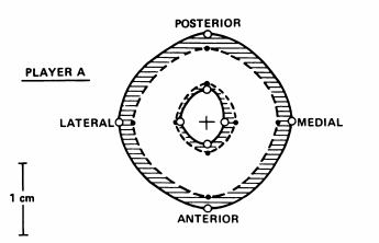
For the 2nd study, we find that the study was on tennis players, not baseball pitchers. The study was done on both tennis players males and females. The men were on average 27 years old and the females were 24 years old. The average number of years the men have been playing was 18 years while the females were 14 years. The study was only on the humerus, not the ulna or the metacarpals. Roentgenograms were used to study the bone changes. The results showed that the humerus of the tennis players all showed hypertrophy, but the increase in size was in width. The thickness of the upper arm bone increased and the intermedullary bone cavity shrunk from the cortical bone increasing in thickness inward. For all 4 parameters used in analysis and recording, the posterior side, anterior side, the medial side, and the lateral side, which basically means that all 4 sides of the cardinal directions were measured.

What is nice is that the 2nd study did cite the first study I have listed and showed the King and associates found that the humerus of the baseball pitchers of the dominant arm were thicker than the arm that was not primarily used for pitching. Others things the 2nd study reveals is that other researcher found that the bone density in athletes for their distal femoral ends was higher than average people .Antoher group of researchers would find that the bone density in cross country runners was also higher with more trabecular bone. The researchers in the 2nd study would conclude by stating that exercise does induce bone hypertrophy.

As for the 3rd study, I post the abstract below…

Abstract

This contribution addresses the following questions: Does unilateral sports-specific strain affect the skeletal system of the athlete? Specifically, can any differences be found in longitudinal growth of the bones of the fore arm and hand in professional tennis players between the stroke arm and the contralateral arm? An investi gation was conducted involving 20 high-ranking profes sional tennis players (12 male and eight female players) between 13 and 26 years of age as well as 12 controls of the same age range. The radiologic examinations of the bones of the forearm and hand yielded an increase in density of bone substance and bone diameter as well as length in the stroke arm as compared with the contralateral arm. Whereas the first results confirm previous findings, the stimulation of longitudinal growth has never been reported. This change in bone structure and size can be attributed to two factors: mechanical stimulation and hyperemia of the constantly strained extremity. It may thus be regarded as a biopositive adaptation process.

Me: What we are finding is that the researchers are now looking at the longitudinal/length difference in the athletes, this time being tennis players. Full article is not available without me paying for it so I was not able to find the full study details. The key thing I can reveal from the study is that in the abstract, the sentence…”The radiologic examinations of the bones of the forearm and hand yielded an increase in density of bone substance and bone diameter as well as length in the stroke arm as compared with the contralateral arm.

It does seem that at least from this 3rd study, the researchers have found that the forearm of the stroking arm to be longer than the contralateral arm.

This shows that the results indicate that there is indeed a bone density and bone thickness increase, but only again anecdotal evidence that the arm with more exercise and loading inducement would cause hypertrophy. So I would try to find 3 more studies to see what the evidence is…

1. Dimensions and estimated mechanical characteristics of the humerus after long-term tennis loading. – Author: Haapasalo H, Sievanen H, Kannus P, Heinonen A, Oja P, Vuori I

2. Effect of long-term impact-loading on mass, size, and estimated strength of humerus and radius of female racquet-sports players: a peripheral quantitative computed tomography study between young and old starters and controls. – Author: Kontulainen S, Sievänen H, Kannus P, Pasanen M, Vuori I.

3. Exercise-induced bone gain is due to enlargement in bone size without a change in volumetric bone density: a peripheral quantitative computed tomography study of the upper arms of male tennis players. – Author: Haapasalo H, Kontulainen S, Sievänen H, Kannus P, Järvinen M, Vuori I.

Analysis & Interpretation: 

From the first study, the researchers did indeed take into account the length of the humerus into measurement. What they did find was that there was indeed a small, but still noticeable increase in player’s humerus length. Quoted…”The playing-to-nonplaying or dominant-to-nondominant arm differences in humeral length ranged from +0.2 to +1.4%, the difference being significant in young male players (+1.4%), young female controls (+1.1%), and older female players (+0.7%). When comparing players’ relative side-to-side length differences with those of the controls, no significant differences were found.” They conclude with ” In conclusion, long-term intensive tennis playing, especially if started in childhood or adolescence, clearly increases thehumeral BMC, BMD, and CWT but seems to have only a minor effect on the width of this particular bone.” and “In older players, the relative side-to-side differences are at the same level or only slightly larger than those in their age-matched controls. This suggests that even intense physical loading of a mature bone is only marginally better in increasing the bone mass, bone density, and CWT of the target bone than the normal daily use of the dominant extremity.”

This shows with some real evidence that at least for young males that probably still had their growth plates, intense playing of tennis might have caused a slight increase in bone length. Even in older females, there was still a little bit of humeral difference between the playing arm and the control, but only at around 0.7%.

For the 2nd study, the measure variables for also young female tennis players on their humerus are

  • Bone mineral content (BMC)
  • Total cross-sectional area (TotA) of bone
  • Cross-sectional area of the marrow cavity (CavA) and that of the cortical bone (CoA)
  • Cortical wall thickness (CWT)
  • Volumetric density of the cortical bone (CoD) and trabecular bone (TrD),
  • Torsional bone strength index (BSIt) for the shaft,
  • Compressional bone strength index (BSIc) for the bone end

The length of the bone was never analyzed but the thickness of the cortical bone was again noticed. The explanation was that “the structural adaptation of the humeral shaft to long-term loading seemed to be achieved through periosteal enlargement of the bone cortex“.

For the 3rd study, the researchers again found that the cortical wall increase in thickness for the males but that the overall bone density didn’t seem to increasem much. The volume of the bone did increase but the increase was in width from the inside where the inter-medullary wall cavity decreased

Conclusion:

It would seem that from the 6 studies I have looked at, 2 of the only studies which did look at humerus length did not a small different between the dominantly used arm and the non-dominant arm. Of course both of the studies were done on young developing tennis players, not baseball pitchers.

I would be more willing to guess that the throwing arm of the baseball pitcher may be longer but the most likely reason is that the longer arm could be from the shoulder joint or elbow joint being progressively moved and subjected to pulls. What I have found is that the baseball pitcher has a very high level of suffering elbow injuries.

What is absolutely clear is that the only way to even have a change to see a length difference between the two arms is for the athletes to start very early in life and age in the sport to have any chance of body modification. This is assuming and guessing that any arm length differences is from the use of the arm with the growth plate cartilage still in there. There is a small chance that frequent intense throwing of the base might indeed exert some effect on the thickness of the growth plates in the arms when the male athletes is still developing

What other height increase researchers like Sky tried to do would not have worked with their knee lunges and kicking because they were probably too old and had no cartilage for them to stretch out.


An Alternative Explanation On Why The LSJL May Actually Help People With Closed Growth Plate Increase Height And Grow Taller (Big Breakthrough!)

I have written many posts for the website but this post I feel may be one of the most important and possible breakthrough posts and insights so far in the research. 

In the last two recent posts I had been focused on this experiment done entitled “A PROCEDURE FOR STIMULATION OF LONGITUDINAL GROWTH OF BONE AN EXPERIMENTAL STUDY” which showed that apparently it is possible to increase height by stripping the periosteum in the region of the long bones close to the growth plates in people who still had their growth plates.

In the last two posts I had not been able to figure out any possible theoretical justification on why the experimental idea  would work until only a few minutes ago. The insight I reached is slightly startling and comes in a form of almost epiphany which makes me wonder whether I could be wrong in my thoughts or not. What I plan to  do now is get Minigolf’s/Tyler’s own thoughts on my new proposed explanation on why his idea on Lateral Synovial Joint Loading has eve been abel to even have any results.

In many posts before I had debated on the feasibility and effectiveness of the idea proposed by Tyler from HeightQuest.com. In some posts I had argued that the idea would not be effective with “Evidence That The LSJL Method Or Loading Is Ineffective In Post-Pubertal Adult Humans? (Important)” and “Why LSJL Might Not Work, An Explanation Using Bone Mechanics And Bone Bridge Studies while in other posts I had argued that it had potential of been effective with “New Proposed Height Increase Method Using LSJL Device WIth Chondrocyte Implants And Growth Factor Injections and “Growth Plate Physeal Longitudinal Growth May Actually Overcome Bone Bridges, New Evidence Showing LSJL May Work

This post will be a completely new proposed alternative idea and explanation on why the LSJL method has been only sporadically effective and seem to only work for a small increment in height increase. I have always been in the school of scientists who believe that science must always be proved through real experimental facts and results. That is why I have always felt that ideas and theories that are too theoretical (but still logically consistent) like the string theory or M-Theory one finds in theoretical physicists attempt to reconcile the differing results of quantum theory and general relativity is not a good path to take.

I personally believe that for a decent theory to work, it must first be able to explain the data we find in our experiments. This theory I am proposing which I am excited in today is something which required the knowledge of almost all of the bone parts to take come up with.

It started from the article I have been looking at recently. The idea of periosteum removal for longtitudinal growth increase was unique but it got me wondering. Here is the proposed hypothesis I have been thinking about…

The LSJL method does work because it manages to induce incisions/fractures/ breaks into the long bone’s surface, past the periosteum causing an entire layer of the long bone to be lifted, thus turning the inner cambium proliferating layer of the periosteum to increase in size, thus adding the neccesary surface thickness increase ,thus height increase.

Let’s look at this hypothesis step by step.

1. You take some thing like a clamp or dumbbell and use a dynamic, alternating hitting/ loading motion on it.

2. On the rare clamp session, one does manage to clamp at just the right magnitude and angle to cause a clear crack in the outer bone layer.

3. Remember that the bone has on the outside a layer known as the periosteum. However, what is not well known is that the periosteum is actually a 2 layer part. There is the outer layer which is hard, protective, and non-proliferative. The inner layer of the periosteum is what is known as the cambium layer, (like the cambium tree layer cells) where the cells are proliferative.

4. If we read the Wikipedia article on the perichondrium, which is the layer which protects hyaline cartilage and the growth plates before being vascularized and then going through ossification, and also read the article on the periosteum, we can see that the the outer two layer are very similar. From the article on the periosteum, we would learn that the perichondrium would actually turn into the periosteum from vascularization. This could suggest that the inner layer of the adult mature human with long bones still would have a type of cell which can differentiate.

Note: At this point I have not done the research but I would guess that the cells we find in the inner layer of the adult human’s long bone of the periosteum is a type of stem cell, or at least a progenitor cell which has not been differentiated into bone cells, specifically the osteocytes/osteoblasts yet.

Update 2/5/2013: From the Wikipedia article on the periosteum, the issue for Note has been resolved. From the article, it specifically states…

The fibrous (inner) layer (of the periosteum) contains fibroblasts, while the cambium layer contains progenitor cells that develop into osteoblasts. These osteoblasts are responsible for increasing the width of a long bone and the overall size of the other bone types. After a bone fracture the progenitor cells develop into osteoblasts and chondroblasts, which are essential to the healing process.

5. From the article we had been looking at, the theory to explain why periosteum stripping would work is that the stripping caused the inner periosteum layer to be pushed out of the outer layer of the periosteum, giving it enough flexibility to differentiate into chondrocytes causing the entire upper layer or area of the long bone to be finally allowed to be pushed up.

url-26. I had always had a reservation over the LSJL idea because the theory is that chondrocytes induced inside the epiphysis has enough force from it’s hypertrphy to push in all 3 dimensions of space past the cortical bone holding it together. Remember that the cortical bone is as strong as steel tensile strength. This issue was raised in the post “Why LSJL Might Not Work, An Explanation Using Bone Mechanics And Bone Bridge Studies“. The problem was always that the chondrocytes induced can not push past the bones, especially since the bones are holding the chondrocytes inside intact in all 3 dimensions.

Note: This main issue over the fact that the chondrocytes have no direction to push in was rasised in the post “How Lateral Synovial Joint Loading Works To Increase Height From Non-Distraction: FAQs and Concerns Answered (Guest Post)

The counter-arguement would be that in growing children, the chondrocytes does have to push upwards against the gravitionaly force and weight of the upper body of the person to increase the height. So any chondrocyte accumulation into cartilage would have to be strong, even if it has only 1 dimension or degree of freedom it can move and increase in size in, ie. longitudinally.

7. So we can say that the LSJL loading on certain people who have working inner periosteum cambium progenitor cells caused a fracture deep enough to allow the cells to be not fully, completely enclosed in by the hard bones. This means that the induced chondrocytes finally have 1 degree of freedom they can increase again. The 1 degree of freedom, with the contraint of not allowing the chondrocytes to push the long bones longitudinally to increase in length will finally be removed, means that the bones will have cells turned to chondrocytes can finally increase again.

Note: Just this last month I would write two critical posts “Local Growth Factor Injections In Aged Periosteum Result In Increased Cambium Proliferation And Cartilage Formation (Breakthrough) and “Increase Height And Grow Taller Using Local Subperiosteal Injection Of Growth Factors IGF-1 And TGF-Beta Percutaneously.” looking at the possible way of using the periosteum inner cells as a possible new alternative growth plate source. The study found yesterday shows that the periosteum inner cambium cells may indeed be used as a possible way to increase the long bone longitudinally.

8. Tyler would show from many protein pathway diagrams looking as multiple gene up-regulations that the technique of dynamically loading bone will cause many of the genes that cause chondrogenesis to be up-regulated. I don’t disagree with him on this fact, and at this point I do believe that the LSJL does cause chondrogenesis in some of the progenitor cells. However, I think he may be mistaken on which cells the loading actually have been having an effect on. Instead of the MSCs we have been thinking has been differentiating from the marrow inside the epiphysis, I propose in this post that the actual cells that are been turned into chondrocytes are actually the inner periosteum cells, the cambium cells. The loading like the stripping of the periosteum, causes the entire inner layer of the cambium layer to be able to push up the top layer of the bone easily,

9. This reasoning and thinking would completely resolve why it is that people like Tyler who has been doing it has seen results, but the results stop after say 3-4 cms. You have hit the bone for too much in the same area of the long bone. From too much hitting, the inner periosteum has become vascularized and completely turned to osteoblasts and calcified. From my own observation of the bones I have shown in video been disected which I have uploaded to YouTube HERE, I can say with complete confidence that even after just 1 day in normal room temperature with normal moisture, the articular cartilage that is exposed to air become extremely hardened to the level of bone-like very quickly. I would guess the process or action which caused this is diffusion of water or air molecules causing the cartilage to loose it’s elastic structure and properties.

10. If Tyler and other people who practice it has been getting results, they have essentially destroyed certain areas of the epiphysis in the periosteum which causes appositonal growth (width wise) for physically mature humans. They have managed to get results, but the results are only a 1 time thing. While it is believed that the bone from remodeling has become desensitized (from piezoelectric sensitivities)  to the loading and we need to increase the loading to get the bone to work again, my thinking at this point is that the loading can be done only once, and cause only a certain amount of real height increase.

The issue with this type of post is that only a few real serious height increase researchers would be able to understand that implications of this post but I feel that this post may be one of those which will dramatically change the direction of this website and the direction of the research we are doing.

Implications and how to solve this issue & Increase height:

url-4What I propose now is that instead of only a blunt instrument to hit or rub the epiphysis like Tyler proposed in LSJL, it would be smarter to instead use 2 types of equipment in succession.

  • First, use a rather sharp, edge instrument to cut/fracture the epiphysis on the side edges, creating effectively a circle of microfractures (aka a closed cloop of indents on the long bone ends which is mostly bone, lacking any muscle tissue tissue). The idea of microfractures is something many former height increase researchers have looked at and does have a high possibility to work. It doesn’t have to be very large or strong, just a few light tapping on the epiphysis close to the closed growth plate with a wedge and hammer like the picture on the right (but not that deep into the bone.
  • Note: Remember that for the distraction osteogenesis of the ilizarov external method or limb lengthening method to happen, the surgeons actually do exactly the same thing. For the initial distraction to hammer, they use a chisel/wedge and hammer to create just a small , unifrom fracture. For our purposes, we do something every similar, but a weaker one creating smaller fractures.
  • The addition of the tapping would make the LSJL far more effective in getting results. After one sees results, the location of the tapping will have to be moved, further and further up (distally) up the distal tibial end. This is because after the induced microfracture of the periosteum is made, that region will become ossified into bone eventually, and the region will be dead for any chondrogenesis. To gain more height, the tapping will have to occur where there is still some cambium progenitor cells left, which means tapping closer to the top of the tibia, creating another layer and chance for the periosteum to increase in thickness.

A New Proposed Height Increase And Grow Taller Method From Periosteum Removal (Breakthrough)

In the last post I would reveal that I had found a study done years ago where a group of researchers found that if you strip the periosteum area close to (but not on) the epiphyseal cartilage area, there was a noticeable increase in longitudinal growth of the bone.

What I would suggest then in this post is an idea for increasing height for people who are still growing which I would say is very simple.

What I would suggest is that the individual would try to stimulate the areas close to the growth plates in their body, near the bone area to lead to increased longitudinal growth.

If we remember the application of the LSJL method using the dumbbells, I would suggest actually something very similar. We take a large, rather blunt heavy instrument, and apply a sliding force to the region of the lower leg/tibia in the epiphysis region, which would be close to the physis (growth plate).

While the rate/frequency of the rubbing has not been determined or optimized yet, I propose that the rubbing action of say a heavy, large surfaced dumbbell would be effective enough to possibly strip some of the periosteum level cells and possibly cause some increased growth.

What I have been thinking is to use a type of chisel that one finds from woodworking like a fishtail chisel wurlhich can evenly and accurately reach a more uniform compressive load action on the bone area.

kneeThe area to rub in the knee region is right on the bone area above where the growth plate would be located. Refer to the picture to the left where the arrow is point at. We would be wanting to rub and try to get the periosteum in the bone area above it.

The action I would suggest is to go with a scrapping or up and down motion on the bone above the tibial physis but the motion should be performed across the entire bone region.

If the study “A PROCEDURE FOR STIMULATION OF LONGITUDINAL GROWTH OF BONE” done on rabbits can be translated to humans even slightly, this method for increased longitudinal growth in growing children should work.

However it might be a better idea to move away from sharp tools that might have been traditionally for woodworking purposes and go with even the hard, but large area edge of a textbook to get the same type of movement and stimulating effect. Interesting, this idea was also proposed in the Book “School Of Height” which I had briefly talked about in the previous post “The School Of Height By A.S. Palko Book Arrived, Thanks Kazlina“, but the idea was instead to rub the knee or joint regions with a certain type of material.


A Procedure For Stimulation Of Longitudinal Growth Of Bone By Stripping The Periosteum (Breakthrough)

I recently got an email from Tyler where he showed me a very interesting study done from Iran about the possibility of getting closed growth plates to regenerate back from using trauma. The study is entitled “Growth Plate Reappearance after Closure in Ankle Radiography for Trauma” and he would write his own post about it entitled “Reverse Ossification” on HeightQuest.com. I was very curious at how he found this really interesting study and he reminded me that you can just go to Google Scholar and type in the phrase “longitudinal bone growth“.

So that is what I did. I typed it in and looked at the results I would find. The results are REALLY good and they were very relevant to our research. From one of the results that appear, I was directed to an old study entitled “A PROCEDURE FOR STIMULATION OF LONGITUDINAL GROWTH OF BONE, AN EXPERIMENTAL STUDY” BY Y. K. W1J, M.B., CH.B., PEIPING, CHINA

The entire document is 13 PDF pages for most of the pages are just pictures and tables and for some height increase researchers like me it was a rather easy and quick read.

The researchers would try to experiment on the long bone with a still functional/working epiphyseal plate and try to either manipulate the epiphyseal plate or the bones around it to get the overal growth rate of the epiphysis to increase for people with limb length discrepancies.

Almost all of the ideas the researchers tried did not work in increasing the growth rate and a lot of the ideas actually decreased or stunted the growth rate.

This would include…

I. Insertion of Foreign Material into a Drill Hole Placed Immediately Distal to the Proximal Epiphyseal Cartilage of the Tibia

II. Indirect Interference of Circulation of Bone

III. Ferguson’s Operation-Curettage of Bone Marrow.

IV. Stripping of the Periosteum

While there was an old study from 1033 by Ferguson which suggested that putting a drill through the bone cose to the epiphyseal and then curetting the marrow inside would stimulate the longitudinal growth, the 3rd type of experiment showed no real longitudinal growth. What was seen however was that the 4th type of bone modification did lead to bone lengthening. When the long bones in the tibia and femur are cut open in the rabbits in the anteromedial space, we do see that the resultant long bones seemed to have increased in length faster than in the controls. The tibia is exposed to a longitudinal incision while the femur is given a lateral incision.

From the study…

Table IV shows the measurements of the length of the bones after the preceding experiments. Figures 1 and 2 illustrate examples of the results obtained. Definite longitudinal overgrowth of the operated bone was observed in all instances except in the tibiae of rabbits Nos. 36, 48, and 51. The bones operated upon were seen to be slightly thicker and rougher than those of the control side, but, in spite of this, the periosteum was not particularly adherent.

From the conclusion/summary…

SUMMARY

In these experiments various surgical procedures were employed, with the hope of finding a method for stimulation of the longitudinal growth of bone. The first three groups of experiments (a repetition with minor modifications of the procedures of Meisenbach, Pearse, Ferguson, and others) failed to produce significant increase in the length growth.

By chance, in several animals, we observed that stimulation of the length growth was produced by the simple procedure of loosening or stripping the periosteum from the shaft of the bone. Consequently, this operation was repeated on twenty-two animals, with quite uniform and significant results, as shown in Table IV. With but three exceptions out of twenty-two rabbits, the operated leg showed definite lengthening when compared with the normal leg on the opposite side. Although the amount appeared to be small, it actually represented an increase of from 5 to 15 per cent. over the normal growth of the bone during that period of VOL. XIX, NO. 4, OCTOBER 1937920 Y. K. WU AND L. J. MILTNER time. Observations of the monthly roentgenograms showed that the most active stimulation of length growth of the bones took place during the first three months following the operation. We are unable to give a definite explanation of the factors which, after the periosteum had been stripped, producing this stimulation of the longitudinal growth.

Analysis & Interpretation:

This study I have found is one of those studies that just are very strange and make very little sense. It would seem that if we remove using some blunt instrument to strip away the periosteum it would lead to some longitudinal growth. I would need to go back to the drawing board and look into the science and theory of bone remodeling to figure out why this even happens. Of course we must remember that this experiment was done on young laboratory animals who still have functional growth plates. It could be very easily that extra activity on the bone areas close to the growth plate can stimulate extra growth.


Natural Height Growth January 2013 – Monthly Website Traffic Data Report.

Note: For the report from last month where I gave both the yearly and monthly numbers of the traffic to the website, go to the post “Natural Height Growth December 2012 – Monthly And End Of Year Website Traffic Data Report

One idea for a regular monthly post I have been doing almost since the beginning of the blog are these monthly reports on the traffic. I have been doing them for all the months and this post will be for the month of January in 2013.

Analysis & Interpretation: 

The month of January seems to have been a little slower and I can only assume that the reason the rate of increase in the traffic has been slowing down over time is from the rate of the number of posts being published here decreasing. In the beginning of the blog, There was around 170-180 blog posts put up in just 1 month!! Now the numbers have decreased to only around 30-50 per month.

What we see is that there was almost 34,000 total visits in the month of January, from the 1st day to the 31st day. The average # of people went from around the 850-900 per day mark in December to around 1000 total visitors/day. Around the middle of the month, the website went viral again where the traffic to the website almost increased by 3X from 1000 total visitors/day to almost 3000 total visitors/day. This was written about in the post “Website Traffic Volume Went Through An Extreme Increase And Other Technical Operational Details“. The news stations and national TV networks picking up on the story over the fact that Tanya Angus passed away and I was one of the earliest sites to report on the news and I got it from TheTallestMan.com website.

Sometimes the traffic is below the 1000 mark around the 950 mark, but other days sees the traffic increase to almost 1150 total visitors/day. Something I do see and realize is a problem is that many of the old and regular visitors to the website are not really coming or reading anymore and I wonder what is missing from the website. The website’s growth is indeed slowing down. This is expected since it is harder for larger websites (and larger things in general) to experience the same level of growth that might have been seen when the website was smaller. Going from 100 total visitors/day to 200 total visitors/day is much easier than going from 1000 total visitors/day to 2000. The very fact that a person’s own success can be their own downfall and that is sort of what is happening recently. Much more negative comments and reception have been appearing recently and the website is only getting slower and slower, and harder and harder to edit and change. There is just a lot more information that needs to be edited and changed.

Personal Note: 

On a personal note, I am in a new relationship with someone new so that has been taking up a lot of my time recently. There have been quite a few days when my social and personal obligations have forced me to not update or write for the website but I do find that I am getting a lot of personal satisfaction with this improvement in my life. I sort of realize now that I do my best work and are the most productive when I am single, focused, and slightly angry. That is sort of a hard state to be in at this point.

Screen Shot 2013-02-03 at 6.22.03 AM


Screen Shot 2013-02-03 at 6.22.39 AM

New Proposed Method To Increase Height And Grow Taller Using TGF-Beta1, Hyaluronic Acid, And Autologous Synovial Fluid

Recently while I was doing research on the link between aggrecan and its connection and effect on the cartilage, I came across this study which seems that suggest that by combining the hyaluronic acid with an external source of synovial fluid we can still get chondrogenic differentiation from mesenchymal stem cells in vivo.

JointAfter reading over the abstract a very simple idea came to me and made me wonder whether in vivo cartilage formation would be just easier to perform. If we remember from our research on microfracture surgery on a possible way to increase height, we note that the microfracture surgery was really very simple. You make a hole in the articular cartilage defect region with a awl and the hole will reach the below the initial bone surface to the sub-cortical level and get to the marrow when the MSCs are. The MSCs leak out and that clotting action creates a another cartilage in the defect’s place after about 2-4 months. However that cartilage would be with disorganized fibers known as fibrocartilage.

This may be one of the easiest proposed ways to increase height yet. The study below shows that if you infuse TGF-beta 1 into a culture of explanted MSCs from the marrow of horse, then the culture will under staining to check for proteoglycan and type 2 collagen see high levels. While the hyaluronic acid by itself does little in terms of anabolic chondrocyte formation, the hyaluronic acid with synovial joint fluid does have some chondrogenic ability. What I propose is a 4 step process for height increase.

  1. Take X-rays of the individual’s knee region to figure out where exactly their articular cartilage ends and where the synovial joint begins.
  2. We are targeting the region inside the synovial joint where the hyaluronic acid (as a GAG) and the synovial joint will be.
  3. We make two incisions on the medial and lateral side of the end of the tibia cutting into the joint, so that the synovial fluid can reach into the inside of the epiphysis.
  4. We inject TGF-Beta1 into the epiphysis, where it will be mixed with the HA and the SF. Since the fluid will flow into the marrow all three compounds will help the MCSs inside to go through chondrocyte differentiation.
  5. The way the incision on the bone will be made will be a from the side to give the bone to heal in a certain way that the MSCs produced will create a band of fibrocartilage which will let is easier for the bone to expand longitudinally.
  6. We make another injection of TGF-beta 1 at a high level, arund 200 ng/ mL into the joint itself to stimulate the chondrogenic property of the HA and SF already in the knee. This will also precent any leaking of MSCS out of the induced incision from diluting the effect of the TGF-Beta1.

The full text for the study is available from clicking the link.

Hyaluronic acid and autologous synovial fluid induce chondrogenic differentiation of equine mesenchymal stem cells: a preliminary study

Abstract

Mesenchymalstem cells(MSC) have the potential to differentiate into distinct mesenchymal tissuesincluding cartilage, which suggest these cells as an attractive cell source for cartilage tissue engineering approaches. Our objective was to study the effects of TGF-beta1, hyaluronic acid and synovial fluid on chondrogenic differentiation of equine MSC. For that, bone marrow was aspirated from the tibia of one 18-month-old horse (Haflinger) and MSC were isolated using percoll-density centrifugation. To promote chondrogenesis, MSC were centrifuged to form a micromass and were cultured in a medium containing 10 ng/ml TGF-beta1 or 0.1 mg/ml hyaluronic acid (Hylartil®, Ostenil®) or either 5%,10% or 50% autologous synovial fluid as the chondrogenesis inducing factor. Differentiation along the chondrogenic lineage was documented by type II collagen and proteoglycan expression. MSC induced by TGF-beta1 alone showed the highest proteoglycan expression. Combining TGF-beta1 with hyaluronic acid could not increase the proteoglycan expression. Cultures stimulated by autologous synovial fluid (independent of concentration) and hyaluronic acid demonstrated a pronounced, but lower proteoglycan expression than cultures stimulated by TGF-beta1. The expression of cartilage-specific type II collagen was high and about the same in all stimulated cultures. In summary, hyaluronic acid and autologous synovial fluid induces chondrogenesis of equine mesenchymal stem cells, which encourage tissue engineering applications of MSC in chondral defects, as the natural environment in the joint is favorable for chondrogenic differentiation.

© 2004 Elsevier Ltd. All rights reserved.