Category Archives: Uncategorized

MATN3 and Rosette Nanotube for growth plate regeneration

LSJL does upregulate MATN3.  A rosette is a hexameric disc shaped aggregate.
Here’s what a rosette Nanotube looks like.  The novel aspect of this is that it can be injected as a liquid.

GROWTH PLATE CARTILAGE REPAIR VIA NOVEL MATRILIN3/ROSETTE NANOTUBE HYBRID MATRIX

“Approximately 15% to 30% of all childhood fractures are growth plate fractures. Because the growth plate determines the length and shape of a mature bone, this type of fracture may result in severe growth abnormalities in children. Pathologically, the growth abnormality is caused by the formation of a bony bridge in the injured growth plate cartilage. Currently, the clinical treatment of growth plate fractures includes the surgical removal of the bony bridge and insertion of autologous fat or cartilage tissue into the empty space to discourage bony bridge reformation. Such surgical procedures are invasive and result in unsatisfactory outcomes. In addition, this treatment is only useful after the bony bridge has formed. Our long-term goal is to understand how to prevent bony bridge formation and improve growth plate cartilage regeneration at cellular and molecular levels and develop the first preventive and therapeutic approach for growth plate fracture. Specifically, the primary objective of this proposal is to evaluate the therapeutic effects of a nano-matrix assembled from matrilin-3 (MATN3) and rosette nanotube (RNT) in a preclinical growth plate fracture model. Our central hypothesis is that the MATN3/RNT nano-matrix specifically promotes chondrocyte growth and enhances chondrogenesis of mesenchymal stem cells (MSCs), while it also inhibits vascularization and osteogenesis at the fracture site{these two things may increase growth plate generation especially since this is supposed to be used for growth plate fracture}. This is the cellular basis for such nano-matrix to improve growth plate cartilage regeneration and prevent bony bridge formation. We will test our central hypothesis and achieve the objective of the proposal by pursuing two specific aims: 1) to determine the ability of MATN3/RNT to prevent bony bridge formation; and 2) to determine the ability of MATN3/RNT to deliver growth factors for further improvement of chondrogenesis and growth plate cartilage regeneration. To achieve the two aims, our overall research strategy includes: 1) optimization of the ratio and dose of MATN3/RNT and its ability and bioactivity for loading growth factors in vitro; and 2) determination of the therapeutic efficay of the nano-matrix in our established growth plate fracture model in rats in long term. The proposed research is innovative: 1) biologically, it simultaneously promotes cartilage regeneration and inhibits bony bridge formation; 2) therapeutically, MATN3 and RNT can be injected as a liquid in a minimally invasive manner, and form a nano- matrix at the fracture site; 3) structurally, the nano-matrix concentrates bioactive MATN3 locally at the fracture site as well as binds TGF-β1 and IGF-1 to achieve multi-functional delivery. With the results of the two specific aims, we expect to 1) realize a synergistic strategy to specifically promote chondrogenesis while inhibiting osteogenesis and vascularization; and 2) develop an injectable approach for the localized delivery of cartilage growth factors. These outcomes have an important positive impact in developing novel, perhaps the first, preventive and therapeutic approach for growth plate cartilage repair. ”

Here’s more info about nanotubes:

Helical rosette nanotubes: a more effective orthopaedic implant material

“Due to the nanometric properties of some physiological components of bone, nanomaterials have been proposed as the next generation of improved orthopaedic implant materials. Yet current efforts in the design of orthopaedic materials such as titanium (Ti) are not aimed at tailoring their nanoscale features, which is now believed to be one reason why Ti sometimes fails clinically as a bone implant material. Much effort is thus being dedicated to developing improved bioactive nanometric surfaces and nanomaterials for biospecificity. Helical rosette nanotubes (HRN) are a new class of self-assembled organic nanotubes possessing biologically-inspired nanoscale dimensions. Because of their chemical and structural similarity with naturally-occurring nanostructured constituent components in bone such as collagen and hydroxyapatite, we anticipated that an HRN-coated surface may simulate an environment that bone cells are accustomed to interacting with. The objective of the present in vitro study is therefore to determine the efficacy of HRN as a bone prosthetic material. Results of this study clearly show that both HRN-K1 and HRN-Arg coated Ti displayed enhanced cell adhesion when compared to uncoated Ti. Enhanced cell adhesion was observed even at concentrations as low as 0.005 mg ml−1. These results point towards new possibilities in bone tissue engineering as they serve as a starting point for further mechanistic studies as well as future manipulation of the outer chemistries of HRN to improve the results beyond those presented here. One such effort is the incorporation of peptide sequences on the outer surface of HRN and/or growth factors known to enhance bone functions. “

Weightlifting and stunted growth

Light weight lifting during development may enhance growth.  Metatarsals are feet bones.

Influence of loading on bone growth at the growth plates in immature rat metatarsals

“Growth of different bones in children is facilitated by different mechanisms according to the anatomical site and function of the bone. Longitudinal bone formation in long and short bones occurs in the cartilaginous growth plates located at each end of the growing bone through a process known as endochondral ossification. This growth continues until a child becomes full-grown at which point the growth plate calcifies to solid bone. It is still unclear how mechanical and biological factors affect bone growth. For the purpose of this study, immature rat metatarsals have been subjected to varying number of cycles (1, 5, 10 and 50 cycles) in order to better understand the effect that mechanical loading has on bone growth. This has been done using two consecutive trials. The trends in these trials were analyzed and compared. Specimens subjected to 5 cycles exhibited the most prominent effect of loading over the course of 16 days. The results of the trials reveal that immature bones are sensitive to cyclic compressive loading. The results revealed a potential threshold below which the loading resulted in an increased growth. Furthermore, simulations of longitudinal bone growth using a thermal-structural coupled analysis, with the findings from the experiment, has been performed. The model results in a stress free structure that is comparable to the growth of the experiments to a certain extent. The model also allowed incorporation of the bent growth that is observed in the experiments.”

“The piston was displacement controlled at 0.01 mm/s up to a predefined maximum load of 0.05 N. After reaching the maximum load, the bones were immediately unloaded. The loading sequence was carried out with varied amount of cycles”

“Compressive loading (static and dynamic) initially reduced the growth rate and growth plate height significantly compared to nonloaded specimens. However, continuing the experiment over a longer time period the results between the groups started to level out. Additionally, growth resumption was observed after loading removal for both statically and dynamically loaded specimen “<-so loading reduced growth rate but not “final” bone length

“At the end of the trial, specimens subjected to 5 cycles exhibited an average percentage growth of 190.9% while the specimens subjected to 50 cycles had an average percentage growth of 166.6%. The control bones grew 166.3% on average. ”

“. Their results showed that both static and dynamic compressive loading initially reduced the growth rate significantly compared to nonloaded specimens. However, continuing the experiment over a longer time period the results between the groups started to level out “<-So you need to change the stimulus to keep getting benefits.

So according to this study, compressive loading exercise should at least alter growth rate.

LSJL Update 9-27-2016

Here’s the last LSJL update.  Here’s the feet images from the this time which have had the best results out of what I’ve been clamping:

20160927_083012

The size increase is not due to flattening of the arches as the arch on the right foot actually looks bigger.  You’ll note that the second and third toe look bigger as well.  My wingpsan has increased from 74.5″ to 74.75″ up from the 72.5″ it was before I started LSJL.  It is very difficult however to take a good wingspan picture.

Part of the trouble with LSJL has been slipping when clamping and a possible solution is that rather than clamping the epiphysis of the bone is to clamp the neck of the bone.

20160927_161437

Considering the spillover of the second and third toe growth, I’d say it’s probably more important to generate clamping force than it is to be at the optimal location.  Clamping at the neck of the bone also clamps the muscle as well which result in more fluid flowing into the bone.  Also clamping at the neck of the bone gets closer to the bone marrow and one key conclusion I’ve come to my LSJL research so far is that the cortical bone and the outer periosteum(growth in width is difficult as well as growth in length) inhibit bone growth and inner periosteum and bone marrow stimulate bone growth.  Distraction osteogenesis both gets rid of cortical bone via fracture and stimulates the bone marrow via blood clot.

By continuing to clamp the epiphysis of the bone is likely the reason why my length gains plateaued as whenever I tried to increase the clamping force the clamp would slip off.  By clamping the neck of the bone I can continue to increase the clamping force without having to worry about slippage.  Hopefully, this will allow me to get some leg length increases that I’ll be able to report otherwise I’ll see if I can continue to gain in the feet and that’ll be proof of concept that I can use to gain more resources to establish better clamping technology to gain in the legs.

I bought a new clamp for my fingers as the standard six inch clamp was just too big.

The problem with this is all the holes in the clamp that make clamping uncomfortable.  If neck clamping with the Irwin Quick Grip clamp doesn’t work


I can doing the C-class clamp again but I worry even with clamping the neck of the bone rather than the epiphysis there’ll be too much slipping.

We’ll see what happens in one to two weeks.  And if it doesn’t seem to be working I’ll switch it up.  Considering my foot growth if I don’t observe results in a reasonable time frame then it’s time to switch things up


LSJL Update 9-13-2016

I tried hand clamping but I seemed to plateau with it so I’m back to using the C-class but more intensely than before.  Here is me doing some bones with a C-class clamp.  I’ve been getting some progress with my feet at least but that could be because changes in the feet are more noticeable because my shoes feel more snug.  Here’s the last feet images I took for comparison.  The first image there is actually the before picture.  Also the II phalanxes(toe closest to big toe) seems longer as well which makes sense since I’m clamping close by.

But my feet seemed to go up in size very quickly once I change methodology of using the C-class clamp over the hand clamp.  So if there’s no changes in a week then I will try something different.  Michael thought about using two C-class clamps at once.  Ideally, yes you want to gain height but the feet is where I’m getting results and if my right shoe no longer fits that would be hard to deny proof and I could use that proof to acquire more resources to translate to height increase research.

Since the II toe is growing I’m worried less about a precise clamping location and more about clamping force.  Now it is possible that the feet could be flattening but the big toe is already pretty straight.  Well if I can keep getting results than such minutiae won’t matter.

Here’s pictures of my feet:

20160913_172028

The right toe is bigger.  I’m not to the point where I need to go up a size for my right foot but I’m closer.

Here’s some unilaterally swollen feet:  The bones don’t physically look longer.  So I don’t think it’s swelling making my feet appear longer.

one-swollen-foot

Here’s another unilaterally swollen foot:

ryans-swollen-feet

Here’s another:

another-unilaterally-swollen-foot

macrophages

This study basically explains that macrophages are key to neo-endochondral ossification:

Fracture healing via periosteal callus formation requires macrophages for both initiation and progression of early endochondral ossification.

“The distribution, phenotype, and requirement of macrophages for fracture-associated inflammation and/or early anabolic progression during endochondral callus formation were investigated. A murine femoral fracture model [internally fixed using a flexible plate (MouseFix)] was used to facilitate reproducible fracture reduction. IHC demonstrated that inflammatory macrophages (F4/80(+)Mac-2(+)) were localized with initiating chondrification centers and persisted within granulation tissue at the expanding soft callus front. They were also associated with key events during soft-to-hard callus transition. Resident macrophages (F4/80(+)Mac-2(neg)), including osteal macrophages, predominated in the maturing hard callus. Macrophage Fas-induced apoptosis transgenic mice were used to induce macrophage depletion in vivo in the femoral fracture model. Callus formation was completely abolished when macrophage depletion was initiated at the time of surgery and was significantly reduced when depletion was delayed to coincide with initiation of early anabolic phase. Treatment initiating 5 days after fracture with the pro-macrophage cytokine colony stimulating factor-1 significantly enhanced soft callus formation. The data support that inflammatory macrophages were required for initiation of fracture repair, whereas both inflammatory and resident macrophages promoted anabolic mechanisms during endochondral callus formation. Overall, macrophages make substantive and prolonged contributions to fracture healing and can be targeted as a therapeutic approach for enhancing repair mechanisms. Thus, macrophages represent a viable target for the development of pro-anabolic fracture treatments with a potentially broad therapeutic window.”

“inflammatory macrophages were required for initiation of fracture repair, whereas both inflammatory and resident macrophages promoted anabolic mechanisms during endochondral callus formation.”

“Periosteal endochondral callus formation progresses via four sequential and interdependent phases: inflammation leading to granulation tissue formation, early anabolism (soft cartilaginous callus formation), late anabolism (hard bony callus formation), and remodeling to reinstate the original bone architecture and mechanical strength”

“recruited inflammatory macrophages are derived from blood monocytes and rapidly infiltrate tissues compromised by injury, abnormal function, and/or infection”
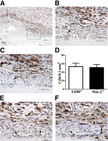
Inflammatory macrophages predominate in fracture granulation tissue and associate with chondrification centers. Representative images of periosteal fracture zone (Supplemental Figure S1A) 7 days after osteotomy and MouseFix plate fixation surgery in 12-week-old C57Bl/6 mice (n = 6 with assessment at multiple sectional depths per sample). All sections were counterstained with hematoxylin. A: Image represents approximately half of the periosteal fracture zone (proximal) with the osteotomy-generated fracture gap (FG) at the bottom left of the image. Granulation tissue predominates at this time point. Tissue section was stained by IHC for the F4/80 pan-macrophage antigen (brown). The black box demarks the region shown in B and C. The blue box demarks the region shown in E and F. B: IHC for F4/80 expression within fracture granulation tissue. The dashed line demarks the interface between the mesenchymal (closest to bone, below dashed line) and inflammatory (above dashed line) stratum. F4/80+ cells with stellate morphological characteristics are evident in both the inflammatory and mesenchymal (arrows) strata. The circle demarks a single osteomac within this field. C: IHC for Mac-2 expression (brown) in a serial section to that shown in B. Arrows point to the same cells indicated in B and highlight the high degree of overlap in F4/80 (B) and Mac-2 (C) staining patterns. The circle indicates the Mac-2neg osteomac identified in B. D: Quantification of the number of F4/80+ and macrophage-like Mac-2+ cells within the mesenchymal stratum of the granulation tissue. An average area of 0.15 mm2 was assessed in six independent samples, and the number of positive cells was not statistically different. E: F4/80+ macrophages (brown, arrows) adjacent to a periosteal chondrification center. F: Mac-2 staining in a serial section to E confirms induction of Mac-2 expression in condensing chondrocyte-like cells within the periosteal chondrification center (blue boxed area). The same F4/80+ macrophages noted in E can be traced and express Mac-2 (arrows). Dashed line in B, C, E, and F demarks the mesenchymal (lower)- inflammation (upper) strata junction within the granulation tissue. Original magnifications: ×10 (A), ×40 (B and C); ×60 (E and F). Scale bars: 100 μm (A); 50 μm (B and C); 37.5 μm (E and F).”

“chondroblasts were identified as F4/80negMac-2+ condensed mesenchymal cells (Figure 1F). F4/80+Mac-2+ (Figure 1, E and F) inflammatory macrophages were observed adjacent to chondrification centers and associated vascular structures.”

” Cartilage and woven bone formations were absent within the periosteal fracture zone [with no macrophages]”

Macrophages are also present in the soft to hard callus transition although this is not as important for purposes as creating the initial growth plate is the limiting factor.

” inflammatory macrophages [were present] in the mesenchymal stratum of the granulation tissue, including some that were associated with developing chondrification centers”

“The developmental vascular canals were broad invaginations with osteoclasts/chondroclasts at the apical tip, presumably excavating the canal path via matrix degradation, followed by a mixture of mesenchymal cells, osteoclasts/chondroclasts, lysosomal cells, macrophages, and endothelial cells.”

“macrophages are pro-mitogenic toward chondrocytes.”<-macrophages undergo chondrocytes to undergo cell division.

Resting and injury-induced inflamed periosteum contain multiple macrophage subsets that are located at sites of bone growth and regeneration.

“Better understanding of bone growth and regeneration mechanisms within periosteal tissues will improve understanding of bone physiology and pathology. Macrophage contributions to bone biology and repair have been established but specific investigation of periosteal macrophages has not been undertaken. We used an immunohistochemistry approach to characterise macrophages in growing murine bone and within activated periosteum induced in a mouse model of bone injury. Osteal tissue macrophages (osteomacs) and resident macrophages were distributed throughout resting periosteum. Tissues were collected from 4 week old mice and osteomacs were observed intimately associated with sites of periosteal diaphyseal and metaphyseal bone dynamics associated with normal growth. This included F4/80+Mac-2-/low osteomac association with extended tracks of bone formation (modeling) on diphyseal periosteal surfaces. While this recapitulated endosteal osteomac characteristics, there was subtle variance in the morphology and spatial organization of modelling-associated osteomacs, which likely reflects the greater structural complexity of periosteum. We also demonstrated that osteomacs, resident macrophages and inflammatory macrophages (F4/80+Mac-2hi) were associated with the complex bone dynamics occurring within the periosteum at the metaphyseal corticalization zone. These 3 macrophage subsets were also present within activated native periosteum after bone injury across a 9 day time course that spanned the inflammatory through remodeling bone healing phases. This included osteomac association with foci of endochondral ossification within the activated native periosteum. These observations confirm that osteomacs are key components of both osteal tissues, in spite of salient differences between endosteal and periosteal structure and that multiple macrophage subsets are involved in periosteal bone dynamics”

“The periosteum is a specialized connective tissue composed of a vascularized and innervated fibrous membrane that encapsulates bone. It has two layers: an outer fibrous capsule layer containing elastic connective tissue (including Sharpey’s fibres), fibroblasts and blood vessels; and, an inner cambium layer containing capillaries, nerves, pre-osteoblasts/bone lining cells, osteoblasts and undifferentiated mesenchymal stromal/stem cells (also referred to as periosteum-derived progenitor cells)”<-these stem cells could potentially be involved in neo growth plate formation.

“Progenitor cells within the endosteum and periosteum have different potential: endosteal progenitors are restricted to osteoblastic differentiation but periosteal progenitors have osteoblastic and chondrocytic bi-potential.”<-It isn’t necessarily true that endosteal progenitors are restricted to osteoblast differentiation it could be influenced by the microenvironment.

Macrophages in bone fracture healing: Their essential role in endochondral ossification.

“In fracture healing, skeletal and immune system are closely interacting through common cell precursors and molecular mediators. It is thought that the initial inflammatory reaction, which involves migration of macrophages into the fracture area, has a major impact on the long term outcome of bone repair. Interestingly, macrophages reside during all stages of fracture healing. Thus, we hypothesized a critical role for macrophages in the subsequent phases of bone regeneration. This study examined the impact of in vivo induced macrophage reduction, using clodronate liposomes, on the different healing phases of bone repair in a murine model of a standard closed femoral fracture. A reduction in macrophages had no obvious effect on the early fracture healing phase, but resulted in a delayed hard callus formation, thus severely altering endochondral ossification. Clodronate treated animals clearly showed delayed bony consolidation of cartilage and enhanced periosteal bone formation. Therefore, we decided to backtrack macrophage distribution during fracture healing in non-treated mice, focusing on the identification of the M1 and M2 subsets. We observed that M2 macrophages were clearly prevalent during the ossification phase. Therefore enhancement of M2 phenotype in macrophages was investigated as a way to further bone healing. Induction of M2 macrophages through interleukin 4 and 13 significantly enhanced bone formation during the 3week investigation period. These cumulative data illustrate their so far unreported highly important role in endochondral ossification and the necessity of a fine balance in M1/M2 macrophage function, which appears mandatory to fracture healing and successful regeneration.”

The failure of endochondral ossification was macrophage specific and not indirectly related to osteoclasts.  Osteoclasts and macrophages come from the same progenitor cell.

Myxedema

Myxedema increases hydrostatic pressure by resulting in increased deposition of connective tissue elements like hyaluronic acid and GAGS(chondroitin).  Maybe there’s a way to use the pathology of this disease to safely increase the deposition of connective tissue to possibly increase height.

Increases in deposition of this elements can result from scar tissue.  Perhaps the separation in limb lengthening surgery can be thought of as a form of scar.  Increases in Fibroblast levels also could increase accumulation of connective tissue.  It is worth it to note that FGFR3 decreases height.  Maybe an interesting possibility is that FGFR3 reduces circulation Fibroblast levels and decreases hydrostatic pressure and results in a height increase that way. Thyroid hormone is thought to increase these connective tissue elements.

Maybe in pregnancy the elevated thyroid causes the bump in the stomach during pregnancy and possibly causes the increase in shoe size and height.  Being pregnant can increase the size and production of the thyroid.

Actors like Marty Feldman who had graves disease was not tall at 5’7″.

Other famous with Graves:

Missy Elliott-F 5’2″

Rodney Dangerfield-M 5’10”

1st President Bush-M 6’2″

Maggie Smith-F 5’5″

According to this paper Graves’ disease–acceleration of linear growth., Grave’s may cause an acceleration of linear growth but I could not find anymore beyond the title.

Body height and weight of patients with childhood onset and adult onset thyrotoxicosis.<-Thyrotoxisis is another name for hyperthyroidism

“The present study has compared body height and weight of thyrotoxic female patients of childhood onset and adult onset. The body height of 141 out of 143 (99%) adult-onset thyrotoxic patients was within the range of mean +/- 2SD for the age-matched general Japanese female population. On the other hand, in 42 patients with childhood-onset thyrotoxicosis, 6 (14%) had their height being greater than the mean + 2SD of general population, and 34 (81%) were taller than the mean value. In 86 patients with siblings, 42 (49%) were at least 2 cm taller than their sisters, and 26 (30%) were more than 2 cm shorter than their sisters. The body weight of 27 out of 42 (68%) patients younger than 20 years was not decreased but was even greater than the mean value for the age-matched general population. The results indicate that excessive thyroid hormone in vivo enhances body height in humans. The increased body weight in some young patients suggests that enhanced dietary intake due to increased appetite in hyperthyroidism has overcome the energy loss with increased metabolism.”

If you look at figure 1 you get quite a interesting figure that shows that of females with adult onset hyperthyroidism tend to be taller than the mean(this is not a longitudinal study so hyperthyroidism could not be a direct measurement of height increase).  I could not excise the figure out of the study you will have to look at it directly.

46, XY pure gonadal dysgenesis: a case with Graves’ disease and exceptionally tall stature.

“Growth was arrested with height remaining at 187 cm after normalization of the thyroid function by treatment with an antithyroid agent, although follow-up to monitor growth was limited to 3 months. In some cases of gonadal dysgenesis, then, Graves’ disease may contribute to an abnormally tall stature.”

So we see that Grave’s disease has an impact on height but that the affect on height is variable sometimes an increase and sometimes a decrease.  Maybe there’s some other variable like FGFR3 levels that influence the effects on height.

This provides more evidence that hydrostatic pressure influences height but perhaps some other stimuli is needed like CNP as increases Fibroblastic stimuli would result in more FGFR3 stimuli in some cases.  CNP would cancel that out.