Category Archives: Uncategorized

Excess Pituitary Stimulated Growth Hormones Increases Male Genitalia aka Penis Size

Something I read from the reddit/r/short threads recently made me remember something I discovered years ago. It seems that male genitalia, (aka Penis Size) can be altered/increased through excess levels of hormones.

I just don't know what the title might be, just please read it, not because of me.

Whoever the poster was, I am guessing that they are feeling the symptoms of a pituitary related tumor, whether benign or malignant. The tumor is stimulating excess HGH release into their system.

Like they said they are already 26, which suggest that their normal growth plates should be closed. This recent events which are indeed unusual for most people doesn’t seem to surprise me. I had guessed that excess HGH can cause even people with closed growth plates to gain 1 full inch in height, just based on the fact that there are still thin slivers of hyaline cartilage tissue in their vertebrate, tibia, femur, etc. which goes through hypertrophy. When you combine all of the hypertrophy the hyaline articular cartilage locations goes through, the total increase in height would often be around 1 inch.

As for height increase, this guy went from 6′ 2″ to 6′ 3″. His height will most likely not increase any further. However, he stated the other fact. His genitalia increased{Tyler-One thing to note is the penis is considered to be like a hydroskeleton}. When I think about this, it all starts to make sense.

Let’s go back a little.

For the normal, average, heterosexual, American men, the most common insecurity might not be their size, in terms of height/stature, but their size as in their “genitalia”. There are in fact entire internet forums dedicated by young men who actively do exercises to increase the size of their genitalia. Since the forums seem to be quite active, I am guessing that these guys who have that pursuit are slightly more successful in their endeavor than ours.

I had proposed in previous posts the idea that stereotypes have in them kernels of truth. I theorized that the stereotype that African American males have larger than average sized genitalia is true. The reason that is the truth is because I guessed that African Americans have higher than average levels of IGF-1 which goes through their system when they are going through puberty.

Higher levels of IGF-1 suggests higher rates of cardiovascular system related medical conditions later in life (diabetes, prostate cancer) which also has a slight correlation to having an earlier than average puberty. I am not sure, but I think I remember seeing studies which show that African American females (at least in urban locations) goes through puberty around 0.5 years earlier than their Caucasian American counterparts. This gives them a slightly less time to grow, assuming all other factors in their growth progression is the same.

Let’s remember our endocrine system. Pituitary gland derived GH goes into the liver, which converts it to IGF-1 which goes to the chondrocyte area (growth plate) of bones making them longer. We also learned that IGF-1 can be created located in the tissue right next to the growth plate. IGF-1 is created in both areas.

If my theory on the link between African Americans and increased IGF-1 levels during puberty is true, it would explain the increase in tissue (size and number) that is not just in bones.

This guy who posted stated that he noticed himself growing again. The obvious guess is that his pituitary gland has gone into overdrive, secreting GH, going to the lier, converting into IGF-1 and that is what is contributing to making his genitalia larger.

Now, this is not the only way chemically. There are a LOT of other options for men to increase the size of their genitalia. (think Relaxin, Prostaglandin ES , Prostaglandin Alpha F2.). In fact, it seems that taking something as simple as Niacin or Nitric Oxide (found in most GNC stores) would be able to do that.

Here is what most people need to realize about growing tissue in their body. Pituitary Gland derived HGH can grow almost all forms of tissue in the human body except for bones, at least interstitially.

If you have ever seen most people suffering from acromegaly, or maybe gigantism when they were younger, they often have wider than average noses, and have wider than average upper torso. The GH going through their system is expanding the costal cartilage in their sternum and the fibrocartilage in their nose even when they are in their 30s, and 40s, of course in small levels.

In fact, analogous to how men in today’s American society worry about the size of their genitalia, American females are too concerned with their mammary gland organs (breasts).

Too many American females get plastic surgery to make their breasts larger, to supposedly give themselves higher levels of confidence/self-esteem. What they don’t understand is that nature (being the generous “mother” that she is) figures out a way to give females who have smaller than average mammary glands larger ones, once they have gone through the process of pregnancy.

In fact, the actress Mila Kunis went on the Conan O’Brien show recently and talked about her experiences in adjusting to the fact that her mammary glands have increased from pregnancy, and now she needs to wear a bra, which she has not had to do throughout her adult life before. So girls, stop going to the plastic surgeon. Just wait for pregnancy to do it naturally. Sure, there is also the added side effect of increased nipple and aereola size, which is for the baby’s mouth. However, the mammary glands definitely increase in size.

Some people might counter and say that the increase is not permanent. I agree on that point, since based on female menstrual cycles, the breasts’ size increases and decreases based on the monthly cycle. However, pregnancy does cause certain chemicals to go often, which have such a dramatic affect on the female body that often the physiological and anatomical changes are completely permanent.

(I am referring to the fact that many caucasian females who had blonde hair before throughout their entire lives had their hair color changed to becoming brunettes because of the release of excess melanin from pregnancy. In fact, you will find many stories of females who had their hair progressively get darker and darker over time after each pregnancy they go through.)

From pregnancy, women gain these changes in their body

  • Feet size increase – Shoe sizes often go up by 1 full size, permanently. This is due to relaxin.
  • Breast size increase – I suspect at least 25% of all women who have gone through multiple pregnancy would notice an increase in cup/bra size that is permanent.
  • Hair color changes – Most pronounced in caucasian females. Light blonde hair turns progressively darker/more brown after each pregnancy.
  • Body hair changes – Increased levels of hair on arms/legs. Also, hair might sprout up in areas of body which wasn’t there before.
  • Hip size increase – Some medical profession might argue with me and say that certain women can never loss the extra 15 lbs they gain from their first pregnancy. They account hip size increase due to fat deposits that become an extra layer. However, I might argue against this and say that hip size increases can be due to realignment of the pelvic/sacroiliac girdle, causing the pubic symphysis to become stretched out.
  • Height increase – Now, this is the phenomena which is most rare. I had guessed that this only happens to maybe 1 our of every 10,000 women.

Interestingly, just today some other woman commented on a previous post “Another Case Of Pregnancy Causing Woman To Grow Taller And Increase In Height”.

Let me show you guys just how often I get messages to this website to that post about this phenomena.

Pregnancy


Fluid Flow enhances Osteoclast Production

Cortical bone is detrimental to longitudinal bone growth as it is not capable of interstitial growth.  Osteoclasts degrade cortical bone.  Degradation of cortical bone in combination with generation of tissue capable of interstitial growth is key to height growth.  Thus if fluid flow increase osteoclastgenesis this should be beneficial.

SUPRAPHYSIOLOGICAL LOADING INDUCES OSTEOCYTE-MEDIATED  OSTEOCLASTOGENESIS IN A NOVEL IN VITRO MODEL FOR BONE IMPLANT LOOSENING

“We aimed to develop an in vitro model for bone implant loosening, allowing analysis of
biophysical and biological parameters contributing to mechanical instability-induced
osteoclast differentiation and peri-implant bone loss. MLO-Y4-osteocytes were mechanically
stimulated for 1h by fluid shear stress using regimes simulating 1) supraphysiological loading
in the peri-prosthetic interface (2.9+/-2.9Pa, 1Hz, square wave),{This is below the minimum chondregenic range which is about 0.1MPa or 100Pa} 2) physiologic loading in the
cortical bone (0.7+/-0.7Pa, 5Hz, sinusoidal wave), and 3) stress shielding. Cellular
morphological parameters, membrane-bound RANKL expression, gene expression
influencing osteoclast differentiation, nitric oxide release and caspase 3/7-activity were
determined. Either Mouse bone marrow cells were cultured on top of loaded osteocytes or
osteocyte-conditioned medium was added to bone marrow cells. Osteoclast differentiation
was assessed after 6 days. We found that osteocytes subjected to supraphysiological loading
showed similar morphology and caspase 3/7-activity compared to simulated physiological
loading or stress shielding. Supraphysiological stimulation of osteocytes enhanced osteoclast differentiation by 1.9-fold compared to physiological loading when cell-to-cell contact was permitted{this is not a bad thing as degradation of bone could potentially be beneficial for height growh}. In addition, it enhanced the number of osteoclasts using conditioned medium by 1.7-fold, membrane-bound RANKL by 3.3-fold, and nitric oxide production by 3.2-fold. The stimulatory effect of supraphysiological loading on membrane-bound RANKL and nitric oxide production was higher than that achieved by stress shielding.  In conclusion, the in vitro model developed recapitulated the catabolic biological situation in the peri-prosthetic interface during instability that is associated with osteoclast differentiation and enhanced RANKL expression. The model thus provides a platform for pre-clinical testing of pharmacological interventions with potential to stop instability-induced bone implant
loosening.”

“[There’s] a specific role for osteocyte communication with osteoclasts “<-Osteoclasts, the cells that break down and reabsorb bone, stem from monocytes and macrophages rather than osteogenic cells.

“The decoy receptor for RANKL, osteoprotegrin (OPG), is released by osteocytes upon
physiological mechanical loading.

“Supraphysiological loading had a flow velocity that was 5.6-fold higher, and the wall shear stress rate that was 4.0-fold higher compared to physiological loading”

“Sixty minutes of supraphysiological loading on MLO-Y4 osteocytes did not change the
cell area, cell perimeter or cell feret’s diameter compared to physiological loading or stress
shielding”<-it would be beneficial if it did because it would indicate that the stem cells could possibly change in diameter.

Osteoblasts, Osteoclasts, and Osteocytes: Unveiling Their Intimate-Associated Responses to Applied Orthodontic Forces

“Histologically, the skeleton comprises 2 types of bone tissue: cortical (compact) and trabecular (cancellous) bone. The cortical bone makes up 80% of the volume in the adult skeleton and the trabecular bone makes up the remaining 20%. The cortical bone forms a peripheral shell in all kinds of bone.”
“The vertebrae are most rich in trabecular bone”
“Modeling of the bone is a process that changes the size and shape of bone either by bone resorption without subsequent bone formation or bone formation without previous bone resorption.”
“microcracks in bone and subsequent apoptosis in osteocytes will activate lining cells in
the remodeling process to stimulate osteoclast formation and bone resorption of the microdamaged area. During the resorption process, growth factors in bone are released, which attract and activate osteoblasts to form new bone under a canopy of bone lining cells.”
“. Osteoblasts form a 1 cell layer covering all bone surfaces. One function is to synthesize an extracellular matrix, consisting of type I collagen fibers and several other proteins, and to subsequently mineralize this matrix into bone”
“Osteoclasts are the only cells in nature that can degrade mineralized bone tissue and are important for physiological remodeling and modeling processes”

Taking Dramamine Seems To Help Children and Adults Grow Taller

While I was doing chemical research for something else today, specifically on the possibility of starting my own chemistry based cosmetic company (similar to what Jessica Alba did with her Honest company to become a billionaire) I found myself looking into at the active ingredients of many of the most common items in one’s bathroom.

It took maybe 3 hours of checking, scouring, and reading (on Wikipedia) to figure out what exactly were the compounds found in my mouthwash, soap, toothpaste, hand lotion, and sunscreen.

Eventually I came across something that was lying on my bathroom medicine cabinet, Benadryl.

I once remembered a friend of mine telling me that you can knock yourself out by drinking a lot of Vick’s Nyquill (aka cough syrup), which has the active chemical compounds of the following below… (source)

Active ingredients (in each 15 ml TBSP) – Purpose

  • Acetaminophen 325 mg  – Pain reliever/fever reducer
  • Dextromethorphan HBr 15 mg – Cough suppressant
  • Doxylamine succinate 6.25 mg – Antihistamine

Antihistamines are a very common type of drug. So from Vick’s Nyquill, I wanted to know the active ingredient of Benadryl. Does Benadryl have a similar effect as Nyquill, where if you take too much, you will get knocked out?

(Side Fun Fact: Interestingly, people often mistake the idea of chloroform as a drug used to knock people out, but it is actually Ether. Chloroform is talked a lot about in Movies, and shown used in Kidnappings, but Ether would be more effective, since it is faster acting on the human system)

(Side Fun Fact #2: Similarly, there is more than one way to make truth serum, which really just lowers people’s inhibitions and acts as a sedative, making them groggy. The recent news about thieves who steal from tourists using the NIghtshade derived Scopolamine and Atropa belladonna. You can also use Sodium Pentothial (aka Sodium Pentathol.)

So what are the active ingredients in Benadryl, the Anti-Histamine Drug most often used to treat allergies like hay fever?

Well, Benadryl is actually a Brand Name for something else, called Diphenhydramine. In the UK, the active ingredient would be acrivastine and/or cetirizine.

While I was doing more research into the compound Diphenhydramine, It led me to an article on Meclizine, aka Meclozine. Medical professionals knew about the bad side effects of Diphenhydramine for a while, and wanted a type of anti-histamine which did not have that type of drowsiness which comes with it.

In my own searching, I remember writing about Meclozine as a one of the most biggest breakthroughs we have made on this website. Tyler back in 2014 wrote a post entitled Game Changing Breakthrough OTC Height Supplement-Meclozine. I myself also wrote about it entitled Achondroplasia Treatment and Increase Height Using Meclozine aka Meclizine – Great News For Parents!

The science behind it seems to be valid. However, there have been a few sources we found on the internet which suggests the exact opposite. Study of possible correlation between BODY HEIGHT DECREASED and MECLOZINE (MECLOZINE)

This one source however does not take away from the multiple sources which show that Meclizine has bone enhancing abilities.

Google patents – Therapeutic agent for systemic bone disease and use thereof – WO 2014141847 A1

This patent was based on an idea to treat achondroplasia by having as one of the active ingredients Meclizine, which they noted is OTC (Over-The-Counter). These same inventors from Japan, wrote a piece called “Meclozine, Motion Sickness Medicine, Has The Therapeutic Potential In Achondroplasia” – (Masaki Matsushita).

It is interesting that Meclozine (aka Dramamine) has sort of replaced the Benadryl/Diphenhydramine as the type of antihistamine to use now.

It seems that Meclozine is actually so common, and easy to get that you can buy it from Amazon. Before, Tyler Linked to the chemical. Here it is again, for sale from Amazon for around $35. (No affiliate link)

Someone asked Tyler whether taking the Meclozine would work with people with closed growth plates. Tyler said that you should take it with doing LSJL. I will let the readers decide on whether it will work for adults.

However, for young children, this chemical seems to have noticeable effects, especially if you get the compound into your blood, and not through your digestive system (aka orally).

Teriparatide potential height increase tool

Teriparatide Improves Trabecular Osteoporosis but Simultaneously Promotes Ankylosis of the Spine in the Twy Mouse Model for Diffuse Idiopathic Skeletal Hyperostosis.

Full study->teriparitadeboneformation

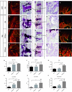
“Diffuse idiopathic skeletal hyperostosis (DISH) is a common skeletal disorder in the elderly, which can develop into periosteal hyperostosis and paradoxically into immobilization-associated trabecular osteoporosis. The bone anabolic agent, teriparatide (TPD), seems to be a rational treatment for the immobilization-associated osteoporosis. However, it can lead to development of hyperostosis lesions in DISH patients. Here, we demonstrate TPD effectively treats trabecular osteoporosis while simultaneously promoting ankylosis of the spine in DISH model tiptoe-walking Yoshimura (twy) mice, compared with the ICR mice. Eighteen male twy mice were divided into three groups, and ICR mice were used as a normal control. Subcutaneous injections of TPD or phosphate-buffered saline (PBS) were performed according to three dosing regimens; 40 µg/kg once daily (TPD × 1 group), 40 µg/kg three times daily (TPD × 3 group), and PBS (control; Ctl group). Treatment was commenced at the age of 7 weeks and continued for 5 weeks. Micro-computed tomography (µCT) and histological analysis were performed. Longitudinal µCT study revealed that trabecular bone volume in both the vertebral body and distal femur decreased with time in the Ctl group, but increased dramatically in the TPD × 3 group. The twy mice developed ankylosis of the spine, the progression of which was accelerated with TPD therapy. We also confirmed that TPD therapy promoted ossification of spinal ligaments. Histomorphometrical study revealed that TPD treatment increased bone formation at the vertebrae enthesis region{This is the key to where this has promise} and in the trabecular bone. TPD therapy effectively treats trabecular osteoporosis, but potentially promotes ankylosis of the spine in patients with DISH.”

“The twy mice are mutant mice showing multiple osteochondral lesions, and have been used as a model for DISH and ossification of the posterior longitudinal ligament”

Teriparatide increased ectopic calcification(Fig3C).

“both TPD × 1 and TPD × 3 treatments promoted ectopic calcification. Histologically, there were no cells inside the ectopic calcification but there were fibroblast- or osteoblast-like cells around the ectopic calcification.”<-Fibroblast cells could be precursors to chondrogenic cells.

Here you can see the effects of teraparatide, it can increase height of the spine but also can cause scoliosis:


teriparatide effects

Study with potential implications to LSJL

TISSUE MODIFICATION OF THE LATERAL COMPARTMENT OF THE TIBIO-FEMORAL JOINT FOLLOWING IN VIVO VARUS LOADING IN THE RAT

“This study describes the first application of a varus loading device (VLD) to the rat hind limb to study the role of sustained altered compressive loading and its relationship to the initiation of degenerative changes to the tibio-femoral joint. The VLD applies decreased compressive load to the lateral compartment and increased compressive load to the medial compartment of the tibio-femoral joint in a controlled manner.

Mature rats were randomized into one of three groups: unoperated control, 0% (sham) or 80% body weight (BW). Devices were attached to animal’s leg to deliver altered loads of 0% and 80% BW to the experimental knee for 12 weeks. Compartment-specific material properties of the tibial cartilage and subchondral bone were determined using indentation tests. Articular cartilage, calcified cartilage and subchondral bone thicknesses, articular cartilage cellularity, and degeneration score were determined histologically.

Joint tissues were sensitive to 12 weeks of decreased compressive loading in the lateral compartment with articular cartilage thickness decreased in the peripheral region, subchondral bone thickness increased, and cellularity of the midline region decreased in the 80% BW group as compared to the 0% BW group. The medial compartment revealed trends for diminished cellularity and aggregate modulus with increased loading.

The rat-VLD model provides a new system to evaluate altered quantified levels of chronic in vivo loading without disruption of the joint capsule while maintaining full use of the knee. These results reveal a greater sensitivity of tissue parameters to decreased loading versus increased loading of 80% BW for 12 weeks in the rat. This model will allow future mechanistic studies that focus on the initiation and progression of degenerative changes with increased exposure in both magnitude and time to altered compressive loads.”

” The VLD applies altered loads in addition to the normal loads across the joint without disruption of the joint capsule while maintaining full use and range of motion of the joint.”

The mice were 9-month old sprague dawley.  The rats had the plates attached surgically so it’s not really an exact approximation of LSJL.  And the 0% BW results were drastically different from control so the apparatus definitely had an effect.
loaded rats

“Gross observation of the tibial plateau revealed minimal erosion or fibrillation of the articular cartilage in all experimental groups.”

“The thickness of the articular cartilage in the peripheral region of the lateral compartment decreased 19% in the 80% BW group as compared to the 0% BW group and 26% as compared to the control group. In contrst, the thickness of the subchondral bone increased 38% in the 80% BW group as compared to the 0% BW group”<-So loading increases subchondral bone “thickness” but does this thickness add to height?

Here’s a comparison between sham control(0% BW) and 80% BW.  Loading definitely decreased chondrocyte number but it didn’t seem to decrease articular cartilage height and subchondral bone height seemed to be higher.
loading on chondrocytes

Subchondral bone thickness is considered height.

Height-Not all HGH and IGF-1

Short and tall stature: a new paradigm emerges.
Full Study->jeffreybaron study

“In the past, the growth hormone (GH)-insulin-like growth factor 1 (IGF-1) axis was often considered to be the main system that regulated childhood growth and, therefore, determined short stature and tall stature. However, findings have now revealed that the GH-IGF-1 axis is just one of many regulatory systems that control chondrogenesis in the growth plate, which is the biological process that drives height gain. Consequently, normal growth in children depends not only on GH and IGF-1 but also on multiple hormones, paracrine factors, extracellular matrix molecules and intracellular proteins that regulate the activity of growth plate chondrocytes. Mutations in the genes that encode many of these local proteins cause short stature or tall stature. Similarly, genome-wide association studies have revealed that the normal variation in height seems to be largely due to genes outside the GH-IGF-1 axis that affect growth at the growth plate through a wide variety of mechanisms. These findings point to a new conceptual framework for understanding short and tall stature that is centred not on two particular hormones but rather on the growth plate, which is the structure responsible for height gain.”

Regulation of growth plate function.Note extracellular fluid is listed as a factor and extracellular fluid flow can be modified by LSJL.

This page lists genes that cause mutations to linear growth.  This page lists other mutations and their effects on stature.

“the vast majority of children with short stature do not have a well-substantiated defect in the GH–IGF-1 axis.”

“Many new genes have been identified that, when mutated, result in short stature or tall stature, the majority of which do not participate in the GH–IGF-1 system”

“Estrogen has complex effects on the growth plate, not only altering growth rate, but also accelerating the loss of progenitor cells in the resting zone and thereby speeding up the developmental program of growth plate senescence, which causes early cessation of growth”

“tumour necrosis factor, IL-1β and IL-6 act directly on growth plate cartilage to suppress bone growth”

“fairly low doses of ionizing radiation, such as a single dose of 10 Gy, can impair longitudinal growth. Mechanical compression across the growth plate also impairs the elongation of bones, which is partly due to decreased enlargement of hypertrophic chondrocytes.”<-Chondrocyte hypertrophy size increases due to lsjl.

“FGFR-3 signalling negatively regulates growth by decreasing proliferation in the proliferative zone, decreasing production of the extracellular matrix, accelerating the onset of hypertrophic differentiation and decreasing the size of the hypertrophic chondrocytes”

“~2% of children who present with idiopathic short stature have mutations in NPR2, Conversely, overexpression of CNP or activating mutations in NPR2 result in tall stature.”

” Binding of CNP to NPR2 stimulates the guanylyl cyclase activity of the receptor, thereby increasing synthesis of cGMP, which activates the type II cGMP-dependent protein kinase”

“Of individuals presenting with idiopathic short stature, 2–15% have mutations in SHOX, with the exact percentage depending on the study. Conversely, increased copies of SHOX are associated with tall stature in individuals with Klinefelter syndrome and other types of sex chromosome aneuploidy”

“Sotos syndrome (characterized by tall stature) is associated with decreased activity of the Ras–MAPK pathway.”

“Ras, a small GTPase, signals via MAPK cascades to phosphorylate numerous cytoplasmic and nuclear proteins, regulating cell proliferation and differentiation.”

“heterozygous mutations in DNA methyltransferase 3A (DNMT3A) cause tall stature, a distinctive facial appearance and intellectual disability.”

“heterozygous mutations in EZH2, which encodes an enzyme that specifically methylates lysine residue 27 of histone 3 (H3K27, which is associated with transcriptional repression), are associated with Weaver syndrome (characterized by prenatal and postnatal overgrowth and a markedly advanced bone age).”

There’s a lot more covered in the study than I mentioned here.  The full study is worthwhile to read.